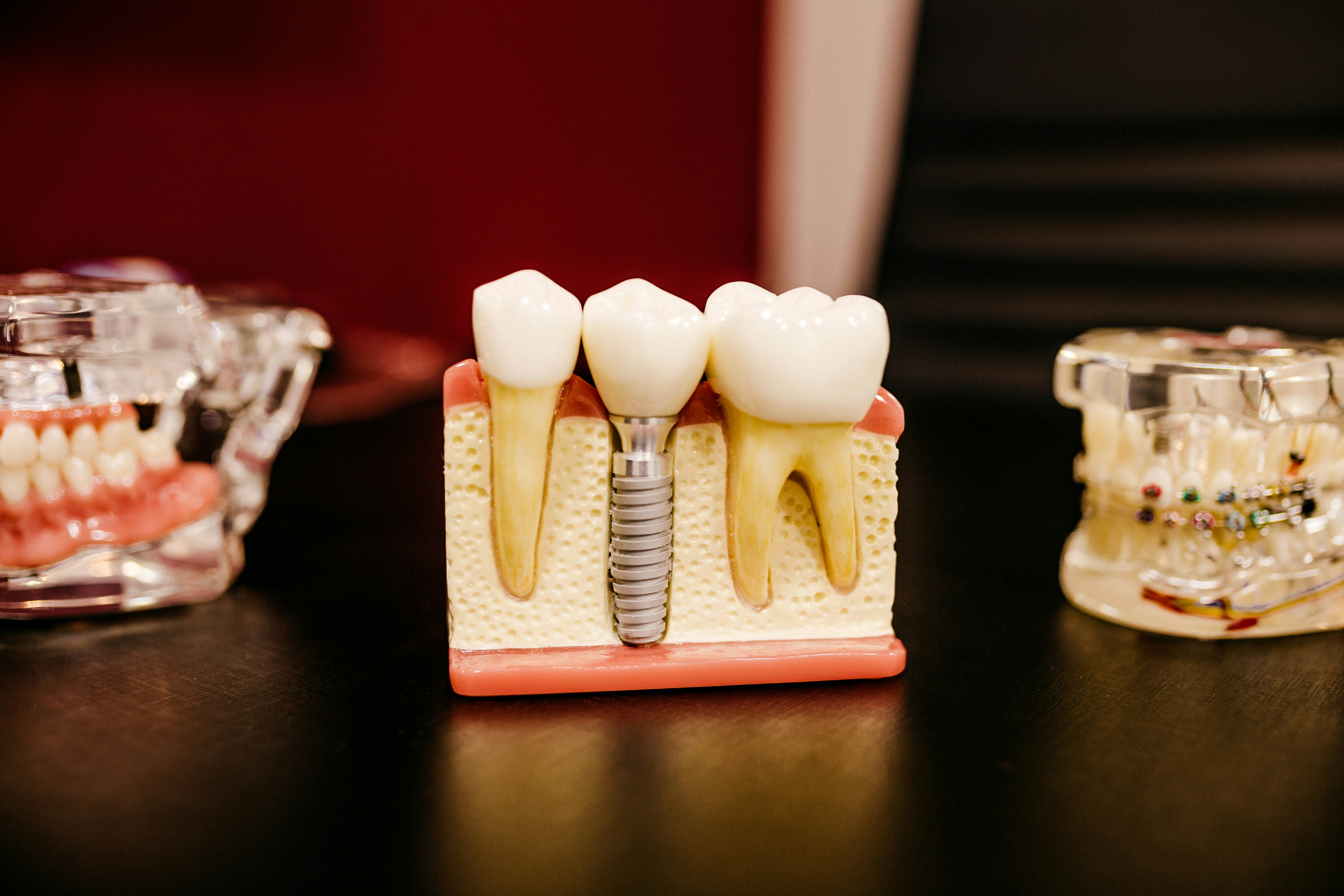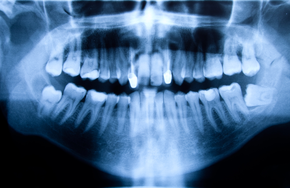
The removal of impacted third molars is one of the most commonly performed surgical procedures. Since the first predictive model of surgical difficulty in 1976 a number of studies to predict surgical difficulty have been performed.
The aim of this review was to determine the patient, radiological, and operative variables associated with surgical difficulty in the extraction of third molars.
Methods
Searches were undertaken in the PubMed/Medline, Scopus and Cochrane Library databases with no restrictions on date or language. Randomised (RCTs) and non-randomised clinical trials (nRCTs) and prospective cohort studies (PCS) were considered. Two reviewers independently selected studies, data was abstracted, and risk of bias assessed using the Cochrane tool for RCTs and the Newcastle-Ottawa Scale (NOS) for cohort studies.
The main outcome variable was surgical difficulty as measured on a VAS by the surgeon (that included the surgeon’s profile), operative time (in minutes), or surgical technique (the need for ostectomy, crown and/or root sectioning). Secondary variables were all characteristics assessed in the included studies that were closely associated with an increase in surgical difficulty. A narrative summary for provided.
Results
- 15 prospective cohort studies involving 2509 patients (3172 lower molars, 528 uppers) were included.
- 2 studies were considered to be of high quality, 10 moderate quality and 3 low quality.
- Almost all of the articles included in used the operative time as the method for estimating surgical difficulty, with some using a 100-mm VAS or the surgical technique in order to rate surgical difficulty.
- Variables investigated
- lower 3rd molars: – age, weight, BMI, mouth opening, cheek flexibility, anxiety level, ethnic background, surgeons experience, impaction depth, available distal space, molar angulation, root morphology, 2nd molar relationship, Inferior alveolar nerve proximity, periodontal space.
- Upper 3rd molars: – BMI, mouth opening, surgeons experience, impaction depth, 2nd molar relationship, root morphology, maxillary sinus proximity, surgical technique complexity.
- For lower 3rd molars
- The majority of variables studied and significantly related to surgical difficulty were radiological. Pell and Gregory type 3C, horizontal or distoangular angulation, the presence of divergent or bulbous roots and tooth germ were considered to increase the surgical difficulty.
- Increased patient age and being overweight were also relevant factors.
- Patient anxiety rated on validated scales directly influenced the difficulty as rated by the surgeon on a 100-mm VAS and increased the operative time.
- 1 study found that difficulty was increased for people of black ethnicity, since the operative time was longer.
- Ostectomy, tooth sectioning, and full bony impaction increased surgical difficulty.
- Surgeon experience was also shown to be a major factor.
- For upper 3rd molars (3 studies)
- Being overweight (as measured by the body mass index) and operative variables such as full bony impaction and surgeon experience also influenced difficulty in terms of operative time and the score on a 100-mm VAS.
- Deep impaction near the maxillary sinus and impaction towards the distal aspect of the second molar were factors associated with increased surgical difficulty.
Conclusions
The authors concluded: –
the following variables were found to be on the spectrum of highly difficult or complex cases: increased patient age and being overweight (patient variables), surgeons with little experience and the use of complex surgical techniques requiring tooth sectioning linked to hard tissue impaction (operative variables), and adverse radiological factors like deep impaction, unfavourable angulation and root morphology, and a close relationship with the second molar, maxillary sinus, or IAN canal.
Comments
The authors have searched 3 major databases with no restrictions on date or language. They considered included a range of study designs with all the included studies being considered prospective cohort designs. 16 different variables were examined in studies involving lower 3rd molars and 8 variables in the 3 studies of upper third molars. The variables considered in each study are helpfully summarised in tables, however only 9 of the variables were considered in 3 or more studies. Consequently, although this review presented a helpful summary of potential factors associated with surgical difficulty the limited data on a number of these variables needs to be taken into consideration.
Links
Primary Paper
Sánchez-Torres A, Soler-Capdevila J, Ustrell-Barral M, Gay-Escoda C. Patient, radiological, and operative factors associated with surgical difficulty in the extraction of third molars: a systematic review. Int J Oral Maxillofac Surg. 2019Nov 14. pii: S0901-5027(19)31357-8. doi: 10.1016/j.ijom.2019.10.009. [Epub ahead of print] Review. PubMed PMID: 31735527.
Other References
Dental Elf – 24th Feb 2017
Panoramic radiography for predicting inferior alveolar nerve injury after third molar surgery