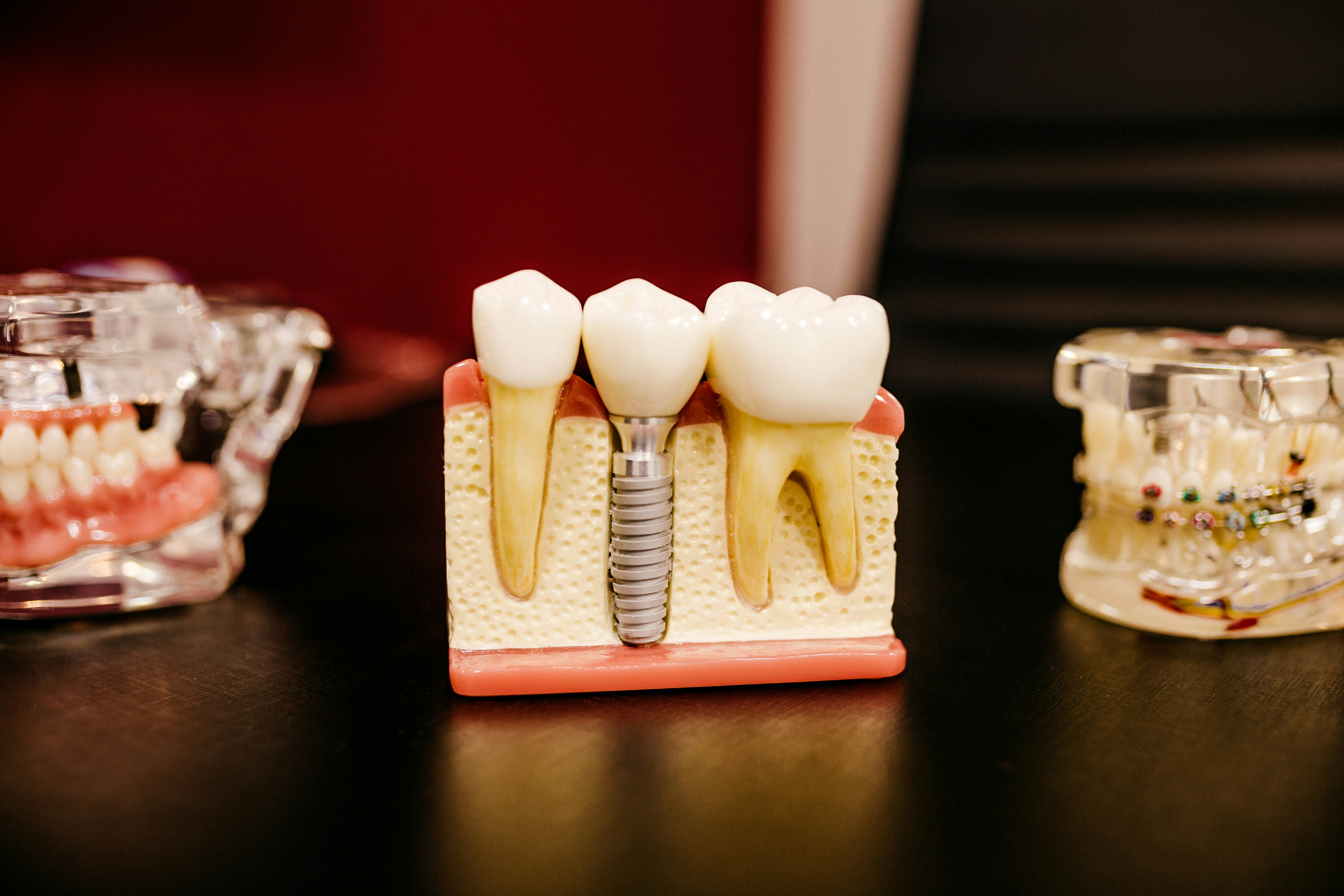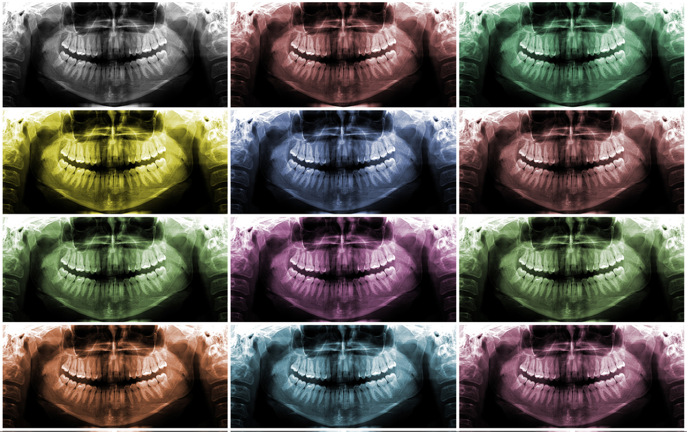
Surgical removal of impacted mandibular third molar (MM3) is a very common surgical procedure. Sensory impairment of the inferior alveolar nerve (IAN) is reported in up to 8% of procedures. Panoramic radiography is commonly used to assess the relationship between the IAN and MM3 and seven radiographic signs have been suggested as predictive of IAN damage. These are, darkening of the root, deflection of the root, narrowing of the root, dark and bifid apex of the root, diversion of the canal, interruption of white line of the canal, and narrowing of the canal.
The aim of this review was to assess the added value of panoramic radiography in predicting postoperative injury of the IAN in the decision-making before MM3 surgery.
Methods
Searches were conducted in the Medline, Embase and World Health Organization International Clinical Trials Registry databases for studies evaluating the diagnostic predictive accuracy of panoramic radiographs for postoperative IAN injury in patients undergoing MM3 surgery. Studies including at least one of the 7 signs of IAN and MM3 juxtaposition reporting postoperative clinical findings and data to calculate False-positive (FP), true-positive (TP), false- negative (FN), and true-negative (TN) proportions for IAN injury were considered.
Two reviewers independently selected studies and abstracted data. Study quality was assessed using the Quality Assessment of Diagnostic Accuracy Studies (QUADAS-2) tool. Positive predictive value (PPV), negative predictive value (NPV), sensitivity, and specificity were recalculated for each included study. Overall pooled estimates of sensitivity, specificity, positive likelihood ratio (LR+), negative likelihood ratio (LR), and diagnostic odds ratio (DOR), with 95% confidence intervals (CIs) using a random effects model. The summary receiver operating characteristic (SROC) curves were also generated.
Results
- 8 studies were included
- 1 studies were considered to have low risk of bias, 7 unclear risk and 1 high risk
- Selected pooled data for the 7 radiographic signs are sown in the table.
| No. of studies | Sensitivity (95% CI) | Specificity (95% CI) | Positive likelihood ratio
(95% CI)
|
Negative likelihood ratio
(95% CI)
|
Diagnostic odds ratio
(95% CI) |
|
| Diversion of Canal | 6 | 0.29
(0.21-0.39) |
0.96
(0.95-0.97) |
5.64
(2.74-11.63)
|
0.81
(0.62-1.07)
|
7.88
(3.04-20.44)
|
| Interruption of White Line of Canal | 8 | 0.39
(0.30-0.48) |
0.84
(0.82-0.85) |
2.67
(1.12-6.38)
|
0.88
(0.63-1.24)
|
3.97
(1.08-14.65)
|
| Narrowing of Canal | 5 | 0.15
(0.07-0.26) |
0.95
(0.94-0.96) |
1.75
(1.09-2.80)
|
0.98
(0.88-1.10)
|
2.51
(1.21-5.21)
|
| Darkening of Root | 7 | 0.49
(0.39-0.60) |
0.81
(0.80-0.83) |
2.40
(1.49-3.88) |
0.66
(0.44-1.01)
|
3.77
(1.54-9.24)
|
| Deflection of Root | 4 | 0.13
(0.05-0.25)
|
0.94
(0.93-0.95)
|
1.67
(0.85-3.27)
|
0.94
(0.85-1.04)
|
1.87
(0.92-3.81)
|
| Narrowing of Root | 5 | 0.06
(0.02-0.16) |
0.97
(0.96-0.98) |
1.61
(0.73-3.56)
|
0.97
(0.90-1.04) |
1.76
(0.77-4.05)
|
| Dark and Bifid Apex of Root | 4 | 0.06
(0.01-0.16) |
0.97
(0.96-0.98) |
1.36
(0.56-3.26)
|
0.98
(0.91-1.05) |
1.44
(0.58-3.59)
|
Conclusions
The authors concluded
For all 7 signs, the added value of panoramic radiography is too low to consider it appropriate for ruling out postoperative IAN in the decision-making before MM3 surgery. The added value of panoramic radiography for determining the presence of diversion of the canal, interruption of the white line of the canal, and darkening of the root can be considered sufficient for ruling in the risk of postoperative IAN injury in the decision-making before MM3 surgery.
Comments
An earlier review of panoramic radiography for IAN injury prediction by Liu et al (Dental Elf – 11th Jun 2015) included 9 studies only 3 of which are included in the current review. That study focused on the darkening of the roots sign and estimated a pooled diagnostic odds ratio for that sign of 6.49 (95%CI;2.92 – 14.44). In contrast this review provides pooled estimates for each of the 7 signs. The 3 signs with the highest diagnostic odds ratios, diversion of the canal, interruption of the white line of the canal, or darkening of the root where considered to have greatest value in ruling in the risk of an IAN injury.
In their discussion, the authors consider the issue of whether the signs are mutually correlated and as such potentially bias the findings. They also highlight some important heterogeneity amongst the studies and that the quality of the included studies was low.
Links
Primary paper
Su N, van Wijk A, Berkhout E, Sanderink G, De Lange J, Wang H, van der Heijden GJ. Predictive Value of Panoramic Radiography for Injury of Inferior Alveolar Nerve After Mandibular Third Molar Surgery. J Oral Maxillofac Surg. 2016 Dec 15. pii: S0278-2391(16)31251-4. doi: 10.1016/j.joms.2016.12.013. [Epub ahead of print] PubMed PMID: 28041843.
Other references
Dental Elf – 11th Jun 2015
Does panoramic radiography predict nerve injury after third molar extraction?