The growth of computer-aided design and computer-aided manufacturing (CAD-CAM) technologies in dentistry is leading to a replacement of conventional impression taking with digital scanning technologies as mentioned in previous blogs.
The aim of this review was to evaluate the scanning and impression times and the radiographic marginal bone loss over time associated with digital scans and conventional impressions for complete arch implant-supported fixed prostheses.
Methods
A protocol was registered on the PROSPERO database. Searches were conducted in the Medline/PubMed, Embase, Scopus, and Web of Science databases with no language or date restrictions. Randomised controlled trials (RCTs) reporting scan and impression times and radiographic marginal bone loss in digital scans and conventional impressions for complete arch implant-supported prosthesis construction were considered. Two reviewers independently selected studies and extracted data. Study quality was assessed suing the Cochrane Risk of Bias Tool for RCTs (RoB 2) and certainty of evidence using the grading of re- commendations assessment, development, and evaluation (GRADE) approach. The scan and impression times and marginal bone loss were assessed using random effects meta-analysis.
Results
- 6 studies published between 2016 and 2022 were included.
- All studies were based in a university setting, 3 studies were published in Italy, 2 in Spain and one in Egypt.
- One RCT was considered to be at low risk of bias with 5 RCTs having some concerns.
- The number of participants ranged from 12 to 56, number of prostheses from 21 to 56 and the number of implants from 87 to 300.
- Follow up times range from 12 – 48 months and implant survival rates from 97.9 to 100%.
- Digital scan time was significantly shorted than conventional impression, MD = 10.1mins (95%CI: 7.46 to 12.55) [3 studies – low certainty evidence].
- There was no difference in marginal bone level between digital and conventional impressions at 6, 12 or 24 months.
Conclusions
The authors concluded: –
Digital scans significantly reduced the time required compared with conventional impressions for complete arch implant-supported prostheses. Nevertheless, additional studies with more consistent methodologies are needed for confirmation. No significant differences were found in radiographic marginal bone loss between treatments performed with digital scans and conventional impressions.
Comments
The authors preregistered their review protocol and searched 4 major databases. Only RCTs were included and only a small number of small studies met the inclusion criteria. As the authors pointed out the clinical procedures varied across the studies with differences in surgical and prosthetic approaches. There were also variations in the evaluation methods with only one study reporting on outcomes at longer than 24 months. While the findings indicate a reduction in clinical times using digital scans compared with conventional impressions the available studies are small with only one study being at low risk of bias. Consequently, the certainty of the evidence is rated as low so findings should be interpreted cautiously.
Links
Primary Paper
Reis INRD, Chamma-Wedemann CN, Silva IAO, Spin-Neto R, Sesma N, da Silva EVF. Clinical outcomes of digital scans versus conventional impressions for implant-supported fixed complete arch prostheses: A systematic review and meta-analysis. J Prosthet Dent. 2023 Oct 19:S0022-3913(23)00640-6. doi: 10.1016/j.prosdent.2023.09.023. Epub ahead of print. PMID: 37865553.
Other references
Dental Elf – Digital impression Blogs
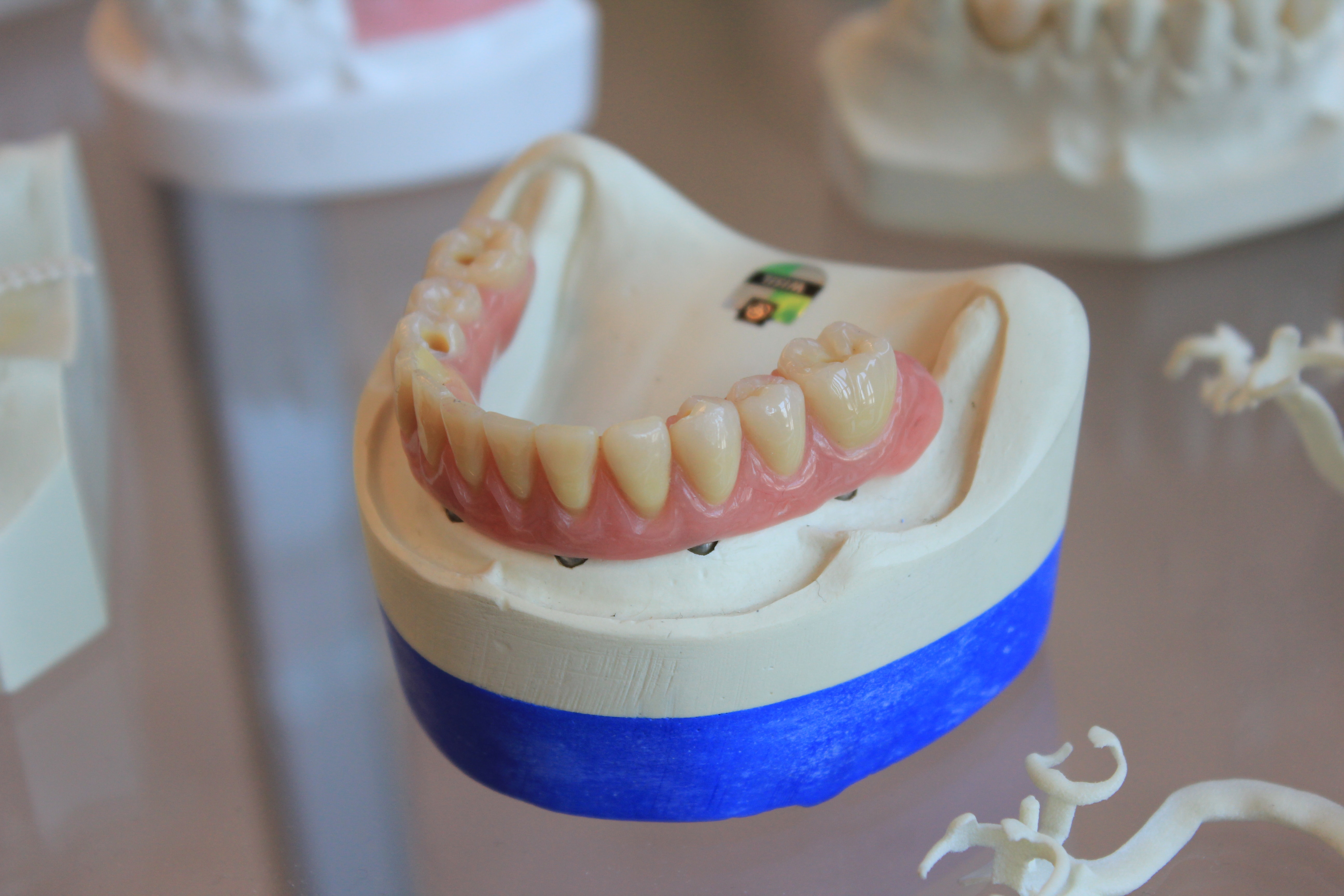
Picture Credits
Photo by Quang Tri NGUYEN on Unsplash