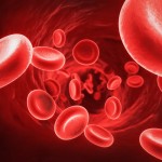
Loss of periodontal structures, cementum, bone, and periodontal ligament (PDL) is a consequence of periodontal disease, and regeneration following successful management is a challenge. Barrier membranes, bone substitutes with or without membranes, and root conditioning with chemical agents have been employed to aid regeneration. Platelet-rich plasma (PRP) is another agent that has been used in guided bone regeneration (GBR) and guided tissue regeneration (GTR). The aim of this systematic review was to assess the influence of PRP on the regeneration of periodontal intrabony defects.
Methods
Searches were conducted in the Medline, Embase, Cochrane Central Register of Controlled Trials, and Cochrane Oral Health Group Trials Register databases. Recent editions of the Journal of Dental Research, Journal of Clinical Periodontology, Journal of Periodontology and The International Journal of Periodontics & Restorative Dentistry were also searched.
Prospective clinical trials with 10 or more patients, reporting the radiographic and clinical outcomes of PRP for regeneration of periodontal defects were considered. Study quality was assessed independently by two reviewers using a modified version of the Cochrane randomised control checklist and the CONSORT (Consolidated Standards of Reporting Trials) statement. The primary outcome was probing pocket depth (PPD) with clinical attachment level (CAL) , radiographic bone level (BL), and changes of marginal gingival level (MGL) as secondary outcomes. The pooled weighted mean difference (WMD) and the 95% confidence interval. a random effects meta-analysis was conducted.
Results
- 21 studies involving a total of 565 patients met the inclusion criteria with 18 contributing to the meta-analysis.
- Three of the four periodontal parameters (BL, MGL, and CAL) showed significant differences between the PRP group and the control group, all favouring the PRP group.
| No. of studies in meta-analysis | Weighted mean difference (95%CI) | |
| Probing pocket depth |
14 |
0.55 mm (-0.09 to 1.20 mm) |
| Clinical attachment level |
12 |
0.58 mm (0.24 to 0.91 mm) |
| Marginal gingival level |
16 |
-0.10 mm (-0.19 to -0.01mm) |
| Radiographic bone level |
2 |
0.76 mm (0.21 to 1.31 mm) |
Conclusions
The authors concluded:
High heterogeneity among studies made it difficult to draw any clear conclusions. Nonetheless, within the limitations of this review, PRP may confer some beneficial effects on the clinical and radiographic outcomes for GTR of intrabony defects.
Comments
This review included a broad search of the literature and identified a large number of randomised controlled trials. However, a majority (13 studies) had 24 or fewer patients (range 5-70) in addition the authors note that there was substantial heterogeneity between the studies in terms of the experimental designs, surgical techniques, graft materials, patient populations, follow-up, durations, and types and methods of preparation of the platelet concentrate. The authors also suggest that the small positive differences seen in 3 of the 4 assessed parameters may not be clinically valuable. They are certainly lower than the values found in the 2006 Cochrane review of GTR by Needleman et al. Consequently the results of this review should be interpreted with caution.
Links
Primary paper
Roselló-Camps À, Monje A, Lin GH, Khoshkam V, Chávez-Gatty M, Wang HL, Gargallo-Albiol J, Hernandez-Alfaro F. Platelet-rich plasma for periodontal regeneration in the treatment of intrabony defects: a meta-analysis on prospective clinical trials. Oral Surg Oral Med Oral Pathol Oral Radiol. 2015 Nov;120(5):562-74. doi: 10.1016/j.oooo.2015.06.035. Epub 2015 Jul 8. Review. PubMed PMID: 26453383.
Other references
Needleman I, Worthington HV, Giedrys-Leeper E, Tucker R. Guided tissue regeneration for periodontal infra-bony defects. Cochrane Database of Systematic Reviews 2006, Issue 2. Art. No.: CD001724. DOI: 10.1002/14651858.CD001724.pub2.

Platelet-rich plasma for periodontal defect regeneration? https://t.co/hGLnlmtHuR
Periodontal defect regeneration using platelet-rich plasma
https://t.co/hGLnlmtHuR
Periodontal intrabony defect regeneration using platelet-rich plasma https://t.co/hGLnlmtHuR
Intrabony periodontal defects and platelet-rich plasma
https://t.co/hGLnlmtHuR
Platelet-rich plasma for periodontal intrabony defects? https://t.co/wRTercRFMo
Don’t miss – Periodontal intrabony defect regeneration using platelet-rich plasma https://t.co/hGLnlmtHuR