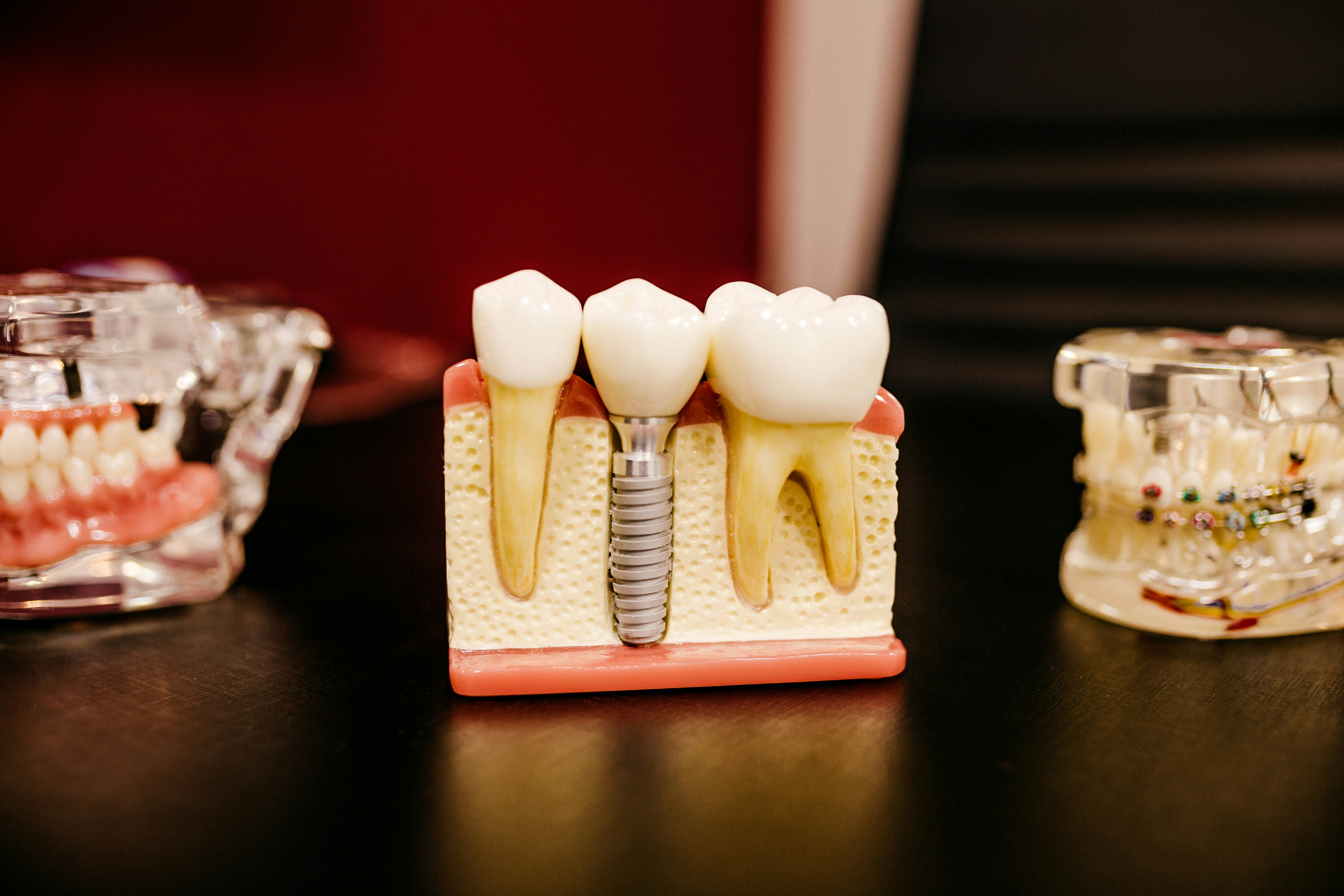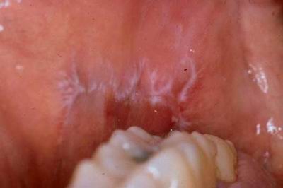
Oral lichen planus (OLP) is a common, chronic inflammatory disorder associated with cell-mediated immune dysfunction. Clinically, it can form bullous, reticular, erosive, atrophic, popular and plaque-like lesions. A range of therapeutic approaches are available that aim to reduce symptoms and manifestation of the lesions (Dental Elf – 22nd Nov 2020). Photodynamic therapy (PDT) is a minimally invasive treatment option for precancerous lesions, cancers of the skin, digestive tract as well as tumours and vascular malformations. It involves three main components; oxygen, a photosensitizer and specific wavelength of visible light. The photosensitizer is activated by the light and causes a series of reactions resulting in irreversible damage and death of the abnormal cells (Chen et al, 2019).
The aim of this systematic review and meta-analyses was to assess the efficacy of PDT in the treatment of OLP and compare this with the efficacy of steroid therapy.
Methods
The Preferred Reporting Items for Systematic Review and Meta-Analyses (PRISMA) were used to inform the methodology of this review. Medline, Web of Sciences, the Cochrane Library, Embase and EBSCO were the databases searched. Studies were included If they assessed the efficacy in the management of OLP. Quality assessment was undertaken through the Cochrane risk of bias assessment tool for randomised control trials (RCTs)and Downs-Black Checklist for non-randomised control trials.
Results
- 418 articles were retrieved across the initial database search. Following removal of duplicates and implementation of the inclusion and exclusion criteria, 16 studies were included for qualitative assessment and from this, 13 for quantitative synthesis.
- The included RCTs had a low or unclear risk of bias. The majority of non-randomised control trials were of high quality.
- 5 types of light sources were utilised in 10 studies and no significant differences (p>0.05) were shown using the random effects model for pooled partial response (the authors reported that as the standard for complete response was strict, half of the included studies achieved no complete response).
- 5 studies used topical application and 5 used gargle administration. Topical application was significantly more effective than the gargle method (p<0.05) and the pooled partial response of topical application reached 0.85 (95%CI: 80 – 0.89).
- PDT was regarded as more efficient in the buccal mucosa and/or lips than tongue and/or gingival mucosa however this was not statistically significant.
- The lesion size was recorded in 6 publications before and after PDT. Following PDT, the lesion reduced in size by 1.53cm2.
- Clinical pain symptoms were measured by the visual analogue scale (VAS) in 6 studies. The VAS score decreased by 3.82 (95%CI: 2.80 -4 .85). Subgroup analysis determined that the efficacy of the diode laser was better than that of the metal halide lamp and LED in relieving pain (p<0.05).
- 5 RCTs with 139 lesions were included to compare PDT with corticosteroids. The efficacy of PDT was better than that of topical corticosteroids (pooled odds ratio: 6.15, 95%CI: 1.65 – 22.97) based on partial response. With the Thongprasom Sign score (method of scoring OLP based on the presence and extent of white striae, erythema and atrophy) (Gobbo et al, 2017), the pooled mean difference was 0.62 (95%CI: -0.46 – 1.71) indicating that the two treatments had similar efficacy to decrease lesion size. The pooled mean difference of VAS was -0.30 (95%CI: -1.99-4.40) indicating a similar improvement of pain between PDT and topical corticosteroids.
- The wavelength of 630-660nm and energy density of 80-150 J/cm2 were commonly used. The duration of irradiation ranged between 120s and 600s.
Conclusions
The authors concluded:-
…PDT could be a valuable optional treatment for the management of OLP… These results indicated that the curative effect of PDT is similar to that of topical corticosteroids in the treatment of OLP.
Comments
The authors have produced a well conducted meta-analysis and systematic review of photodynamic therapy for oral lichen planus. There were limited numbers of studies in the sub-group analyses however the comprehensive data extraction protocol and statistical calculations provided clarity on which studies were pooled for each parameter and the significance of findings. The results of this study suggest that photodynamic therapy may offer patients with oral lichen planus improved pain relief and better efficacy compared to topical corticosteroids. However, as the authors highlighted, both photodynamic therapy and topical corticosteroids had a similar efficacy to decrease lesion size. There were no reported outcomes for malignant transformation. Whilst no changes in clinical practice can be recommended, the study does highlight a potential future avenue for treatment.
Links
Primary Paper
He, Y., Deng, J., Zhao, Y. et al. Efficacy evaluation of photodynamic therapy for oral lichen planus: a systematic review and meta-analysis. BMC Oral Health 20, 302 (2020). https://doi.org/10.1186/s12903-020-01260-x
Other references
Chen, Q., Dan, H., Tang, F. et al. Photodynamic therapy guidelines for the management of oral leucoplakia. Int J Oral Sci 11, 14 (2019). https://doi.org/10.1038/s41368-019-0047-0
Gobbo M, Rupel K, Zoi V, et al. Scoring systems for Oral Lichen Planus used by differently experienced raters. Med Oral Patol Oral Cir Bucal. 2017;22(5):e562-e571. Published 2017 Sep 1. doi:10.4317/medoral.21833
Dental Elf – 22nd Nov 2017
Picture Credits
By James, Candice, Mai – http://bohone09.wikispaces.com/Group+1, CC BY-SA 3.0,