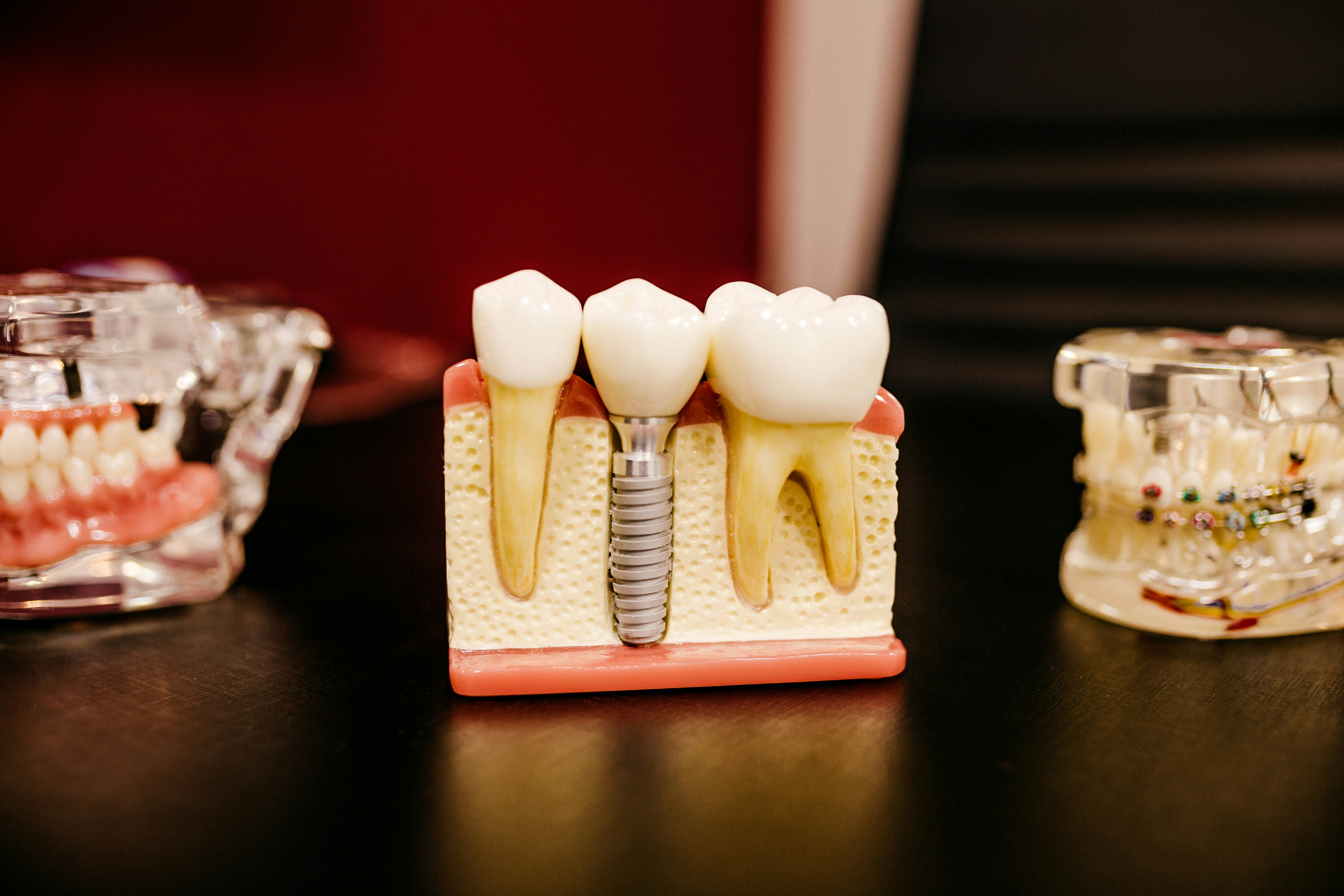
Maxillary sinus floor elevation is commonly performed to create sufficient bone fraction to enable the placement of dental implants. The aim of this review was to assess the bone fraction and implant survival rate and to determine whether the bone fraction is affected by the grafting material or growth factor applied.
The Medline and the Embase databases together with references of identified studies were searched randomized controlled trials (RCTs) or cohort studies where sinus floor elevations with autogenous bone (controls) were compared with autogenous bone combined with growth factors or bone substitutes, or solely with bone substitutes (test groups) were included. Study quality was assessed independently by two reviewers with disagreements being resolved by discussion.
Twelve split mouth RCTs were included. Meta-analysis revealed no significant differences in bone formation after 5 months between the various techniques identified (autogenous bone; autologous bone with growth factors (platelet rich plasma); or autogenous bone and bone substitutes (bovine hydroxyapatite, bioactive glass, corticocancellous pig bone). A significantly higher bone fraction was found in the autogenous bone group compared to the sole use of b-tricalciumphosphate (P = 0.036). At one years the overall implant survival rate showed no significant difference between implants
The authors concluded
Bone substitutes combined with autogenous bone provide a reliable alternative for autogenous bone as sole grafting material to reconstruct maxillary sinus bony deficiencies, for supporting dental implants after 5 months. Adding growth factors (platelet rich plasma) to grafting material and the sole use of b-tricalciumphosphate did not promote bone formation.
Rickert D, Slater JJ, Meijer HJ, Vissink A, Raghoebar GM. Maxillary sinus lift with solely autogenous bone compared to a combination of autogenous bone and growth factors or (solely) bone substitutes. A systematic review. Int J Oral Maxillofac Surg. 2011 Nov 16. [Epub ahead of print]
Related reviews
A Cochrane review ( Esposito et al) published in 2010 looked at, ‘whether and when augmentation of the maxillary sinus are necessary and which are the most effective augmentation techniques for rehabilitating patients with implant-supported prostheses’.
That review included 10 trials and concluded at that time: –
Conclusions are based on few small trials, with short follow-up, and judged to be at high risk of bias. Therefore conclusions should be viewed as preliminary and interpreted with great caution. It is still unclear when sinus lift procedures are needed. 5 mm short implants can be successfully loaded in maxillary bone with a residual height of 4 to 6 mm but their long-term prognosis is unknown. Elevating the sinus lining in presence of 1 to 5 mm of residual bone height without the addition of a bone graft may be sufficient to regenerate new bone to allow rehabilitation with implant-supported prostheses. Bone substitutes might be successfully used as replacements for autogenous bone. If the residual alveolar bone height is 3 to 6 mm a crestal approach to lift the sinus lining, to place 8 mm implants may lead to fewer complications than a lateral window approach, to place implants at least 10 mm long. There is no evidence that PRP treatment improves the clinical outcome of sinus lift procedures with autogenous bone or bone substitutes.
Esposito M, Grusovin MG, Rees J, Karasoulos D, Felice P, Alissa R, Worthington HV, Coulthard P. Interventions for replacing missing teeth: augmentation procedures of the maxillary sinus. Cochrane Database of Systematic Reviews 2010, Issue 3. Art. No.: CD008397. DOI: 10.1002/14651858.CD008397.