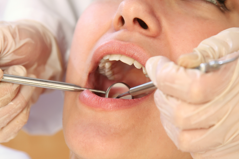
Yesterday we looked at the management of large carious lesions and today we are considering the other end of the caries process, the detection of the early or non-cavitated carious lesion (NCCLs). With changes in the severity of dental disease in some population there has been more interest in the identification and management of the early stages of the disease process (NCCLs) when the process can be arrested or enamel remineralised. The aim of this review was to evaluate the sensitivity, specificity and reliability of detection systems for NCCLs.
The Medline, Embase, Cochrane Oral Health Group’s Specialized Register, Cochrane Central Register of Controlled Trials and SCIELO databases were searched together with hand search of the Caries Research Journal table of contents.
In-vivo or in-vitro studies of natural early enamel of dentine carious lesions validated against gold standards that provided sensitivity and specificity data or allowed this to be calculated were included. Study quality was assessed using an 11-element scale.
- Forty two studies yielding 85 separate assessments were included. Methods evaluated were; Visual (V), Caries Lesion Activity Assessment (CLAA), Laser Fluorescence (LF) [including DIAGNOdent (DD) and Quantitative Light-induced Fluorescence (QLF)], Radiographic (R), Fibre-Optic Trans-Illumination (FOTI), and Electrical Conductance (EC).
- Overall, the quality of evidence on detection methods was rated ‘poor’, except for EC that was rated ‘fair’.
- The majority of studies using visual criteria were conducted in vitro as were those involving radiography and DIAGNOdent, electrical conductance and FOTI.
- The table shows a summary of the performance measures for all detection systems (sound versus non-cavitated and cavitated lesions)
[table id=40 /]
The authors concluded
There is a large variation in sensitivity and specificity values for methods and a lack of consistency in definition of disease and analytical methods. EC and QLF seem to be promising for detection of early lesions. For both cost and practicality considerations, visual methods should remain the standard for clinical assessment in dental practice.
Note:-This study was sponsored by a research grant from Colgate Palmolive Company. Prof Roger Ellwood is an employee of the Colgate Palmolive Company.
Comment
The QUADAS tool may have been more appropriate instrument to have measured the quality of the included studies rather than the 11-item scale used here. Data is present for both sound verses cavitated and non-cavitated lesions in the review and the and there are minor differences between the sensitivity ranges presented in the abstract to the tables in the main text. While it is understandable that the majority of studies were undertaken in-vitro this also needs to be taken into account when considering transferring this information to the clinical situation.
Links
Gomez J, Tellez M, Pretty IA, Ellwood RP, Ismail AI. Non-cavitated carious lesions detection methods: a systematic review. Community Dent Oral Epidemiol 2013; 41: 55–73. © 2012 John Wiley & Sons A/S