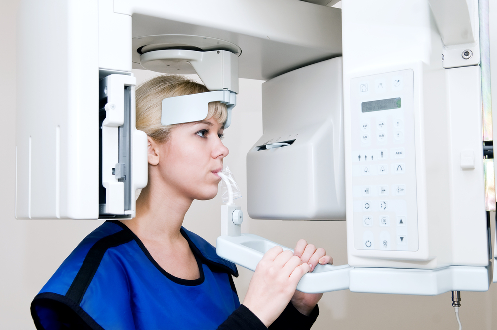
After third molars maxillary canine are the most common tooth to suffer from impaction or ectopic eruption, with a prevalence ranging from 1-3%. Localisation of the impacted tooth is needed to assess treatment options and predict the difficulty and duration of treatment. While conventional 2D radiographs are still the main diagnostic aid the use of 3D imaging is increasing. Although conventional radiographs can suffer with anatomic superimposition and geometric distortion cone-beam computed tomography (CBCT) uses higher radiation dosages.
The main aim of this review was to compare the diagnostic accuracy of cone-beam computed tomography (CBCT) and conventional radiography (CR) in the localisation of maxillary impacted canines.
Methods
Searches were conducted in the PubMed, CINHAL, Web of Science, Cochrane Library, Pro-Quest Dissertation Abstracts and Thesis database and Google Scholar databases.
Studies comparing CBCT imaging with conventional 2D radiographs (panoramic, periapical, occlusal, and cephalograms) in maxillary impacted canine localisation were considered.
Two reviewers independently selected studies, abstracted data and assess study quality. Case-control and cohort studies were assessed using the Newcastle-Ottawa Scale and the Quality Assessment of Diagnostic Accuracy Studies (QUADAS-2 ) tool was used for diagnostic accuracy studies.
Results
- 8 studies were included.
- There were 2 diagnostic accuracy studies and 6 cross-sectional studies
- The accuracy of CBCT ranged from 50% to 95%, and the accuracy of conventional radiographs ranged from 39% to 85%.
- Inter-observer agreement was higher for CBCT than conventional radiographs.
Conclusions
The authors concluded: –
- CBCT is more accurate than conventional radiographs in localizing maxillary impacted canine.
- CBCT may be more reliable than conventional radiographs
- There is no robust evidence to support using CBCT as a first-line imaging method for impacted maxillary canine evaluation
Comments
This review pulls together the data on 8 studies comparing CBCT and conventional radiography for the diagnosis of impacted maxillary canine. Only 2 of the included studies were considered to be diagnostic accuracy studies and no data on sensitivities and specificities are provided in the main paper. Two of the included studies did not involve patients. The sample sizes for the included studies involving patients ranged from 18-60 and the authors highlight that there was an obvious spectrum bias in selection of the included patients.
The author indicates that CBCT is more accurate than conventional radiography. However, that accuracy comes at the cost of increased radiation dosage. Current recommendations from both the British and American Orthodontic Societies recommend that CBCT may be indicated for localising the assessment of an impacted tooth when the information cannot be obtained adequately by lower-dose conventional radiography. Before using CBCT or any diagnostic test clinicians should be asking, What will I do if this test is positive ?, What will I do if this test is negative? If neither result would change you treatment plan, don’t order the test.
Links
Primary paper
Eslami E, Barkhordar H, Abramovitch K, Kim J, Masoud MI. Cone-beam computed tomography vs conventional radiography in visualization of maxillary impacted-canine localization: A systematic review of comparative studies. Am J Orthod Dentofacial Orthop. 2017 Feb;151(2):248-258. doi:10.1016/j.ajodo.2016.07.018. Review. PubMed PMID: 28153153
Other references
Kevin O’Brien’s Blog on this review