
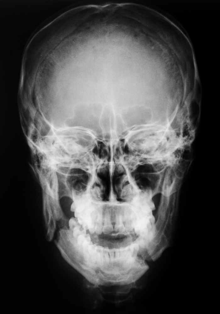
The prevalence of mandibular fractures among facial fractures is high, reaching 76% of all facial fractures. The mandibular regions that fracture most frequently are the mandibular condyles (56.5%), mandibular symphysis (45.0%), body (25.5%), and angle (16.5%). The locations of mandibular fractures are related directly to the fragility of these bone areas: the condyle is the mandibular area with the lowest bone thickness and is most frequently fractured.
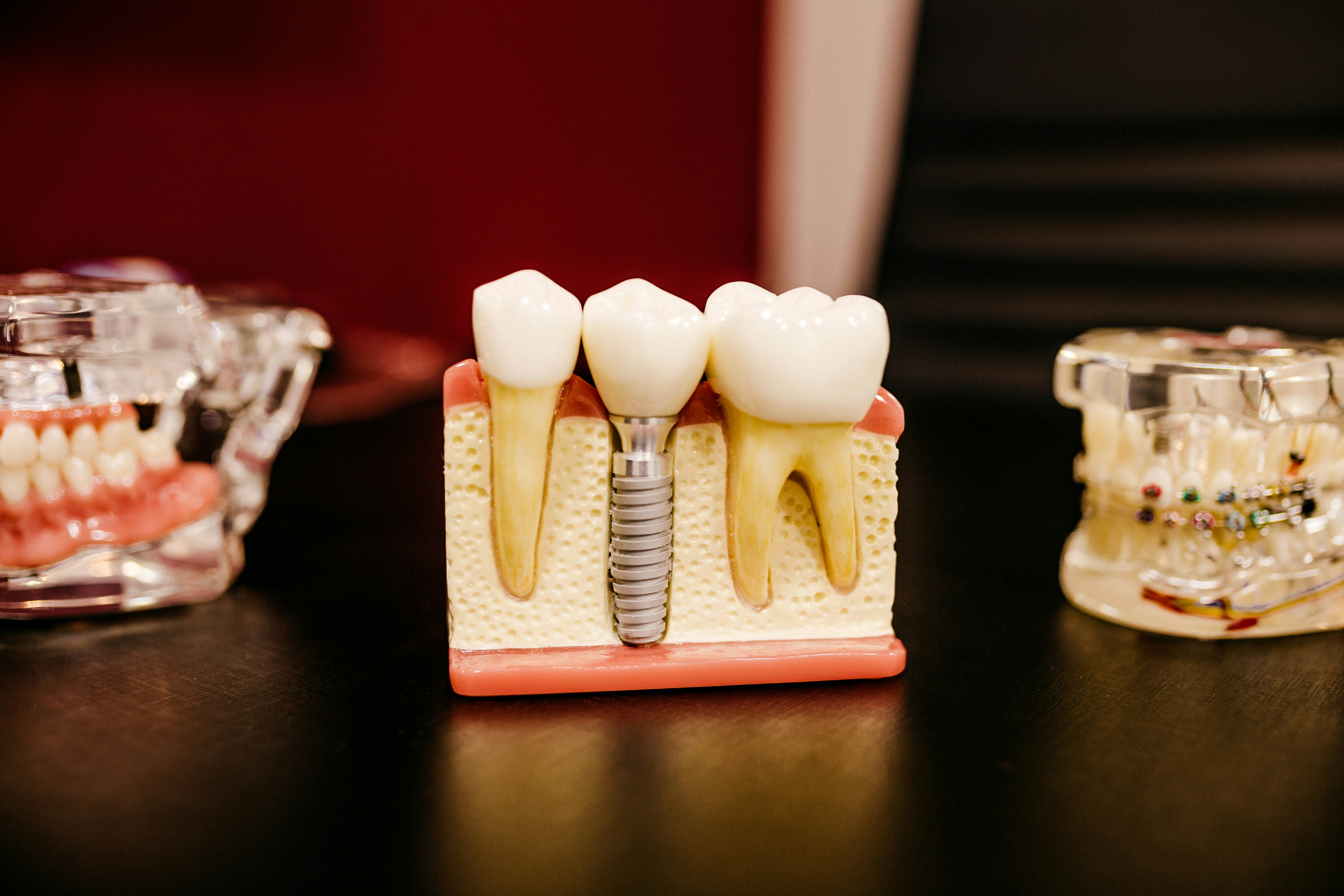
The third molar, when present, may generate a weak area in the mandibular angle and predispose this region to fracture. The authors wished to investigate this relationship.
Methods
This systematic review and meta-analysis was performed according to the Preferred Reporting Items for Systematic Reviews and Meta-Analyses (PRISMA) statement.
The population comprised patients with mandibular fractures; the exposure was the presence or absence of a third molar (3M) and the different positions of the third molar; the comparison was with other mandibular fractures; the outcome was an angle fracture.
The following inclusion criteria were applied in this systematic review: observational studies (cross-sectional, case– control, and prospective and retrospective cohort studies. Exclusion criteria were iatrogenic jaw fracture from surgery and fracture in patients with diseases of bone metabolism (osteopenia and osteoporosis).
The electronic survey was conducted by two authors in the PubMed, Scopus, Web of Science, Cochrane Library, and VHL (Virtual Health Library; BIREME (PAHO/WHO)) databases, and included publications up to January 2016, without language restriction.
The assessment of the quality of the studies included was performed using the Newcastle–Ottawa scale (NOS) for case-control studies and a modified NOS for cross-sectional studies.
Results
The systematic Search found 35 papers out of 704 which fulfilled the inclusion criteria. There were 22 case-control studies and 13 cross-sectional. 28 out of the 35 were used in the meta-analysis.
On the risk of bias assessment 5 case controlled and 2 cross sectional studies scored 8/9 on the Newcastle-Ottawa scale.
Meta-analysis results expressed as an Odds Ratio (OR)
| Odds Ratio | 95% CI | Heterogeneity (I2) | |
| Odds of mandibular fracture (angle) than any other mandibular fracture if 3M’s present. | 3.27 | 2.75-4.16 | 81.3 |
| Mandibular angle fracture in the presence of a 3M | 3.83 | 3.02-4.85 | 83.1 |
| Comparing a Pell and Gregory B v. A+C | 1.44 | 1.06-1.96 | 87.2 |
| Comparing a Pell and Gregory A v. B+C | 0.60 | 0.45-0.81 | 87.1 |
| Comparing a Pell and Gregory C v. A+B | 1.19 | 0.57-2.46 | 96.1 |
Conclusions
The authors concluded: –
The results suggest that the presence of the third molar increases the chance of angle fracture by 3.27 times and that the most favourable positions of the third molar for angle fracture are classes B and II, whilst classes A and I act as protective factors.
Comments
There are three areas that need to be taken into account when considering the authors result and conclusions.
The first point is that angle of jaw fractures are not common. In a recent paper looking at mandibular jaw fractures presenting at a London teaching hospital the approximate annual incidence for angle fracture was 17 in 100,000. About half the individuals present with 2 mandibular fractures and the male: female ratio was 6.6:1(Rashid et al. 2013).
Secondly the cause of mandibular fractures varies with geographical location. Roughly 90% of fractures in the UK data were due to interpersonal violence and in Asia/India the same proportion is due to motor vehicle accidents. Fractures resulting from violence were most commonly associated
with the angle region while those related to road traffic accidents usually involve the condyle, body and parasymphysis (Nasser et al. 2013).
Thirdly the results are expressed as an odds ratio(OR). Even though OR is a legitimate way to express an effect size especially in regard to case-control studies it may not have been the best way to convey the information in this case. When events are rare the difference between odds and risk are small but when events are common (>20%) this difference can become large, in this review it’s about 60%. From the ‘analysis = fracture forest plot in Fig.3’ the OR is 4.15 (i.e. out of 5 jaw fractures 4 will be at the angle) but when this is converted to risk ratio (RR) we get a value of 1.60 (i.e. The presence of a 3M increases the risk by 60%).
Though there is no doubt that the presence of 3M increases the chance of a mandibular angle fracture the results need to be interpreted with caution due to both high confounding and heterogeneity.
Links
Primary paper
Armond ACV, Martins CC, Glória JCR, Galvão EL, Dos Santos CRR, Falci SGM.Influence of third molars in mandibular fractures. Part 1: mandibular angle-ameta-analysis. Int J Oral Maxillofac Surg. 2017 Jun;46(6):716-729. doi:10.1016/j.ijom.2017.02.1264. Epub 2017 Mar 11. Review. PubMed PMID: 28291569.
Other references
Nasser M, Pandis N, Fleming PS, Fedorowicz Z, Ellis E, Ali K. Interventions for the management of mandibular fractures. Cochrane Database Syst Rev. 2013 Jul 8;(7):CD006087. doi:10.1002/14651858.CD006087.pub3. Review. PubMed PMID:23835608.
Rashid A, Eyeson J, Haider D, van Gijn D, Fan K. Incidence and patterns of mandibular fractures during a 5-year period in a London teaching hospital. Br J Oral Maxillofac Surg. 2013 Dec;51(8):794-8. doi: 10.1016/j.bjoms.2013.04.007.Epub 2013 Jun 2. PubMed PMID: 23735734.
Dental Elf – 21st May 2013

Mandibular fractures: the influence of third molars – Thoughts on Life and Dentistry
8 years ago