Changes in hard and soft tissue dimensions are inevitable post-extraction. Preservation of tissue can be beneficial in restorative treatments and aesthetics. Alveolar ridge preservation (ARP) is a proactive treatment option that aims to dimensionally preserve hard and soft tissue post extraction. Numerous biomaterials have been utilized to carry out ARP.
The aim of this review was to evaluate and compare the effects of different ARP techniques and rank their performance.
Methods
The review adhered to the PRISMA guidelines, and the protocol was published in PROSPERO. A literature search was conducted electronically on MEDLINE (PubMed), EMBASE, Cochrane Central Register of Controlled Trials and Scopus. A manual search was also performed on numerous journals like the British Dental Journal, British Journal of Oral and Maxillofacial Surgery, Clinical Implant Dentistry and Related Research. Online registries providing information about in-progress clinical trials were reviewed. Two reviewers independently assessed relevant articles; a third author was consulted where disagreements were present. Interrater reliability (IRR) was consistently maintained >80%.
The review considered only Randomized Control Trials (RCTs) for patients undergoing tooth extraction with or without ARP. ARP involving different bone grafts and membrane biomaterials were reviewed with all possible comparisons including spontaneous healing. Soft tissue outcomes were evaluated and compared including horizontal linear changes, Vertical buccal linear changes and keratinized mucosa thickness (KMT) changes. RCTs with at least 10 patients with minimum of 6 week follow up post extraction were included.
Risk of bias was assessed based on tool reported in the Cochrane Handbook for Systematic reviews of Interventions. Network geometry plots were generated to compare treatment interventions. Contribution plots, inconsistency plots, predictive interval plots, surface under the cumulative ranking curve (SUCRA) and multidimensional scale rankings were used to present results of the NMA. Strength of the evidence was assessed using the GRADE criteria for the NMA.
Results
- 22 studies were included in the qualitative synthesis, 11 in the NMA quantitative synthesis after exclusions.
- 7 were associated with a low risk of bias, 4 with moderate risk, none with high risk.
- KMT measurements were reported in 7 studies. The most common comparison was between non-crosslinked collagen membranes and crosslinked collagen membranes. No statistical difference was observed with reference to the control group.
- Vertical buccal height was reported in 5 studies in the NMA. The most common comparison was between crosslinked collagen membrane and the control group. According to the SUCRA ranking crosslinked collagen membrane was ranked highest by a wide margin.
- Horizontal linear changes were reported by 4 studies considered by the NMA. Autogenous soft tissue group ranked highest in the SUCRA ranking followed by non-crosslinked collagen group.
Conclusions
The authors concluded:
Within their limitations, the findings of the present systematic review and NMA confirmed that the use of crosslinked collagen membranes and autogenous soft tissue grafts, with a minimum of 6-week follow-up, represented the best bio- material choices for sealing sockets during ARP in terms of minimizing post-extraction soft tissue dimensional shrinkage
Comments
The systematic review and NMA have highlighted that there is a variable response in soft tissue dimension change based on the biomaterials used in alveolar ridge preservation. There were a few limitations observed in the review: Heterogeneity in measurement techniques for soft tissue dimensional changes. For example, physical impression materials may compress tissues, resulting in undersized models and scanning might be inaccurate due to contamination with blood or moisture. Follow-up duration for the included studies might be another limitation as it should have been at least 6 months to accommodate further changes to hard and soft tissues during post-extraction healing. Some confounding factors were evident. For example, with regards to smoking status, 8 studies included smokers, 2 did not report smoking status and one considered only non-smokers. Heterogeneity was also noted in techniques for assessing dimensional changes, chosen reference points, studied outcomes, analysis of buccal wall integrity, statistical reporting approach, type of bone filler and use of pre/post operative antibiotics, all of which can potentially influence the results.
The surgical techniques, suturing, incision design and area in question (molars, non-molars) can potentially affect results as well. Lastly the limitations of NMA studies are also to be taken into consideration for example, SUCRA ranking will help a clinician understand the degree of potential positive effects but may not necessarily balance this out with the negative clinical effects. It also does not take into consideration the cost or clinician’s familiarity with the materials.
Overall, the quality of available evidence is limited. The studies included in the NMA were less in number with a small number of comparisons, but the NMA has facilitated indirect comparison of interventions where direct comparisons were not assessed in literature. This also highlights a need for further research in soft tissue dimension changes particularly, comparisons of differences in soft and hard tissue dimension changes which may have direct clinical benefits of restorative treatment planning.
Links
Primary paper
Canullo L, Pesce P, Antonacci D, Ravidà A, Galli M, Khijmatgar S, Tommasato G, Sculean A, Del Fabbro M. Soft tissue dimensional changes after alveolar ridge preservation using different sealing materials: a systematic review and network meta-analysis. Clin Oral Investig. 2022 Jan;26(1):13-39. doi: 10.1007/s00784-021-04192-0. Epub 2021 Oct 20. PMID: 34669038; PMCID: PMC8791918.
Other references
Dental Elf – 28th Apr 2021
Picture Credits
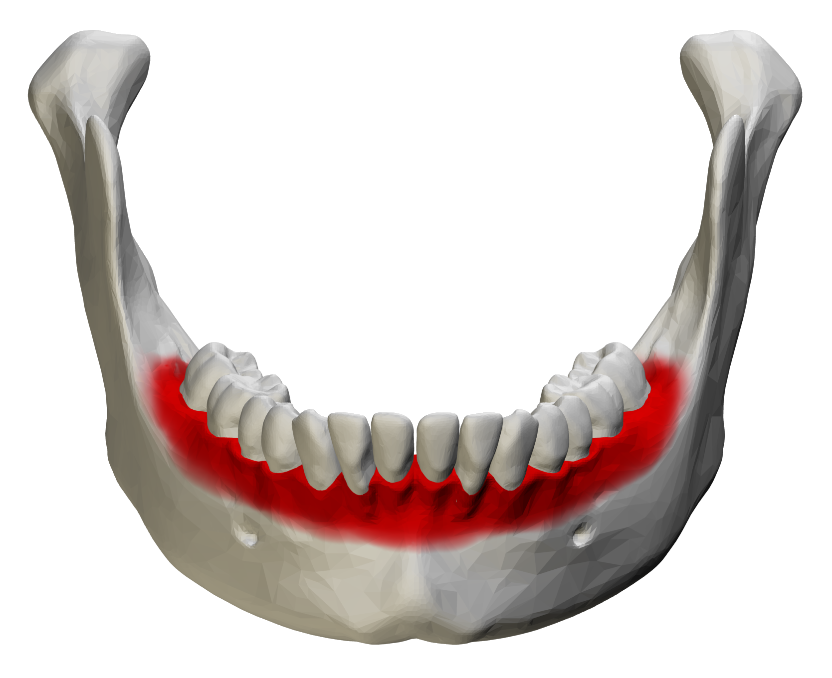
By BodyParts3D/Anatomography – BodyParts3D/Anatomography, CC BY-SA 2.1 jp, https://commons.wikimedia.org/w/index.php?curid=34108339