
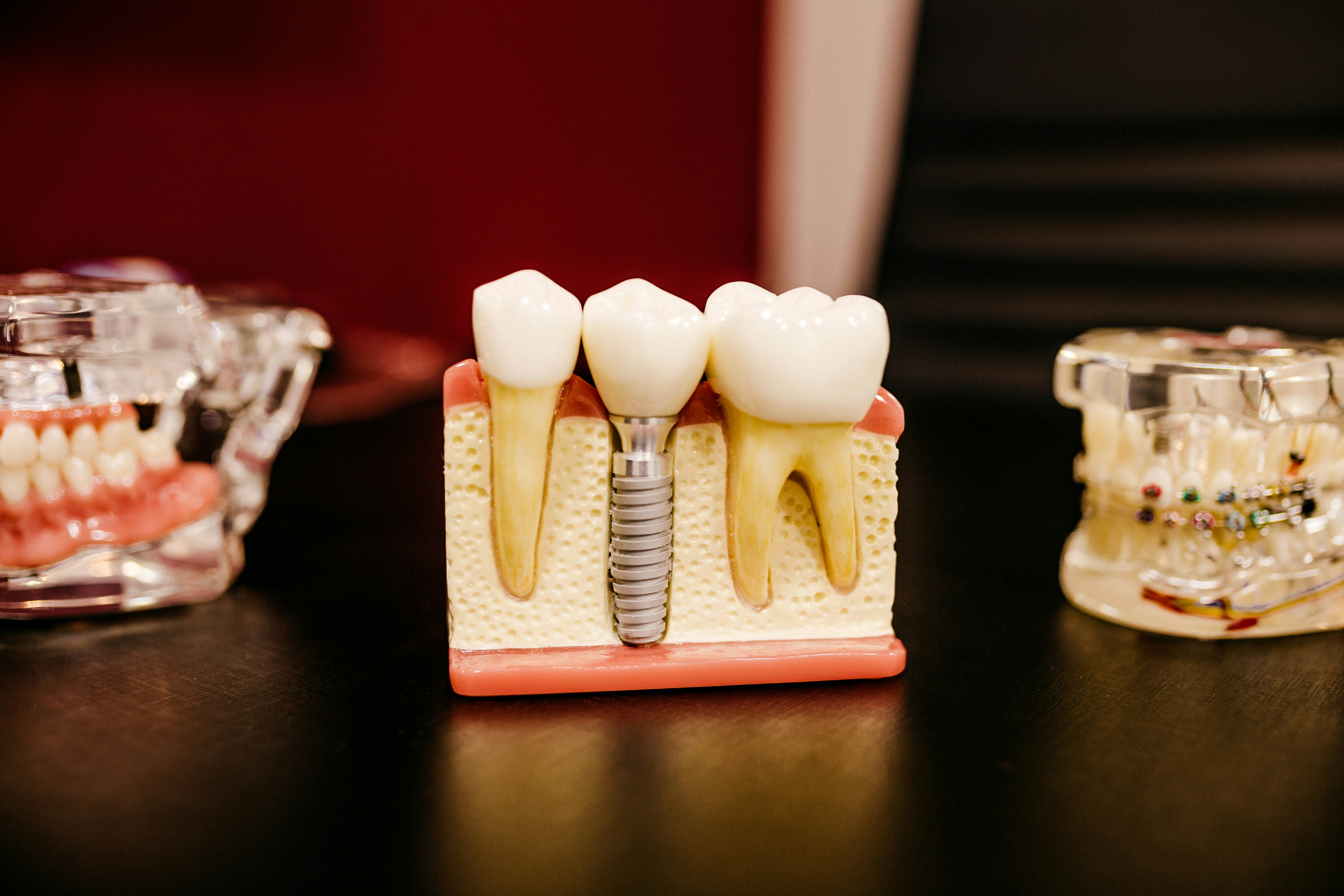
Following tooth loss remodeling of the alveolar bone and soft tissue is a normal physiological response. This can result in a loss of height and width of the alveolar ridge in the order of 40-60%. To reduce this loss a wide range of alveolar ridge preservation (ARP) procedures have been proposed.
The aim of this review was to assess the effects of alveolar ridge preservation on bone and gingival tissue site dimensions, keratinised tissue width, histological bone characteristics and patient-based outcomes.
Methods
Searches were conducted in the Medline, Embase, Cochrane Central Register of Controlled Trials (CENTRAL), and LILACS databases. These were supplemented by hand searches of the reference lists of identified papers and the journals; Clinical Oral Implants Research, Clinical Implant Dentistry and Related Research, European Journal of Oral Implantology, Implant Dentistry, International Journal of Oral and Maxillofacial Implants, International Journal of Periodontics and Restorative Dentistry, Journal of Clinical Periodontology, Journal of Dental Research, Journal of Oral and Maxillofacial Surgery, Journal of Periodontology, Oral Surgery, Oral Medicine, Oral Radiology, Oral Pathology and Endodontics.
Two questions were considered,
1; is there any additional benefit of alveolar ridge preservation techniques over unassisted healing in terms of the following: (i) horizontal and vertical alveolar ridge dimensions, (ii) soft tissue conservancy measured through linear and volumetric analysis, (iii) histological characteristics of the bone, (iv) keratinised tissue dimensions and (V) patient-based outcomes?
- what are the estimated size effects on (i) horizontal and vertical alveolar ridge dimensions, (ii) gingival tissue conservancy measured through linear and volumetric dimensional changes, (iii) histological characteristics of the bone, (iv) keratinised tissue dimensions and (V) patient- based outcomes, following different alveolar ridge preservation techniques?”
Randomised controlled trials (RCTs) and controlled clinical trials (CCTs) with unassisted socket healing as a control group were considered for the main question, while large prospective case series were considered to assess side effects. Two reviewers independently selected studies with a third reviewer adjudicating in disagreements. Study quality was assessed and meta-analysis carried out where possible.
Results
- Of all the included studies only 2 were considered to be at low risk of bias.
- 9 studies (8 RCTs, 1 CCTs) involving a total of 194 patients were included for question one.
- 6 studies were of a parallel design 3 split-mouth design.
- Follow up ranged from 3- 6 months.
- Standardised mean difference (SMD)
- Vertical mid-buccal bone height between ARP and a non-treated site was 0.739 mm (95% CI: 0.332 to 1.147).
- Comparing proximal vertical bone height and horizontal bone width 0.796mm (95% CI: -1.228 to 0.364) and 1.198 mm (95% CI: -0.0374 to 2.433).
- Examination of ARP sites revealed significant variation in vital and trabecular bone percentages and keratinised tissue width and thickness.
- Adverse events were routinely reported, with three papers reporting a high level of complications in the test and control groups and two papers reporting greater risks associated with ARP.
- No studies reported on variables associated with the patient experience in either the test or the control group.
- 37 studies (29 RCTs, 7 CCTs and 1 case series) were included for question two.
- Follow up ranged from 3- 9 months.
- A pooled effect reduction (PER) in mid-buccal alveolar ridge height of -0.467 mm (95% CI: -0.866 to -0.069) was recorded for GBR procedures and -0.157 mm (95% CI: -0.554 to 0.239) for socket grafting.
- A proximal vertical bone height reduction of -0.356 mm (95% CI: -0.490 to -0.222) was recorded for GBR, with a horizontal dimensional reduction of -1.45 mm (95% CI: -1.892 to -1.008) measured following GBR and -1.613 mm (95% CI: -1.989 to -1.238) for socket grafting procedures.
- Five papers reported on histological findings after ARP. Two papers indicated an increase in the width of the keratinised tissue following GBR, with two papers reporting a reduction in the thickness of the keratinised tissue following GBR. Histological examination revealed extensive variations in the treatment protocols and biomaterials materials used to evaluate extraction socket healing. GBR studies reported a variation in total bone formation of 47.9 ± 9.1% to 24.67 ± 15.92%.
- Post-operative complications were reported by 29 papers, with the most common findings soft tissue inflammation and infection.
Conclusions
The authors concluded: –
Within the limitations of this study, the following conclusions can be drawn:
- ARP results in a significant reduction in the vertical bone dimensional change following tooth extraction when compared to unassisted socket healing.
- A reduction in horizontal alveolar bone dimensional change was found when ARP was compared to unassisted socket healing, but the difference between techniques was not found to be statistically significant.
- No evidence was identified to clearly indicate the superior impact of a type of ARP intervention (GBR, socket filler and socket seal) on bone dimensional preservation or keratinised tissue dimensions. Currently, it is not known whether a biomaterial or a treatment protocol is superior to others.
- There is insufficient evidence to demonstrate a difference in the amount of vital bone formation following GBR or socket grafting techniques.
- Inflammation was common following ARP.
- The majority of the studies evaluating ARP procedures presented with high or unclear risk of bias. Clinical recommendations derived from this study should be interpreted with caution.
Comments
This review has undertaken a broad search strategy to address the two detailed questions considered and summarises a significant amount of information. In terms of the main question they have chosen to include both RCTs and CCTs and it is interesting to note that they were able to only include one more CCT than the 2015 Cochrane review by Atieh et al. Also only 1 of the RCTs is common to both reviews. The Cochrane review is more circumspect in its conclusions compared with the current review noting:-
There is limited evidence that ARP techniques may minimise the overall changes in residual ridge height and width six months after extraction
In contrast the current review highlights
a significant reduction in the vertical bone dimensional change following tooth extraction when compared to unassisted socket healing
The current review only includes a small number of studies in relation to the main question and these only involve a small number of patients. The quality of the included studies is also limited and the outcomes are only reported in the short term consequently the findings should be viewed with caution and high quality long-term trials are required. In addition the authors note an number of potential confounders including measurement error, as a number of techniques were used and erros here could have a important bearing on the findings.
Links
Primary paper
MacBeth N, Trullenque-Eriksson A, Donos N, Mardas N. Hard and soft tissue changes following alveolar ridge preservation: a systematic review. Clin Oral Implants Res. 2016 Jul 26. doi: 10.1111/clr.12911. [Epub ahead of print] Review. PubMed PMID: 27458031.
Other references
Cochrane Oral Health Group Blog – Maintaining the jaw bone to support dental implants
Dental Elf -2nd June 2015
Alveolar ridge preservation: limited evidence for current techniques

flyerLPA
9 years agoTheDentalElf
9 years agoAlveolar ridge preservation techniques: evidence limited | Dental News | Tooth Wise
9 years agoTheDentalElf
9 years agoKhalid Waleed Alsahly
9 years agoTiago Marques
9 years agoflyerLPA
9 years agoTheDentalElf
9 years agoTheDentalElf
9 years agoTheDentalElf
9 years agoflyerLPA
9 years ago