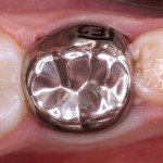
Globally dental caries affects 60-90% of children, most commonly in primary molar teeth. If this is not managed it can lead to pain and infection and impact on ability to grow and thrive.
The aim of this review was to evaluate the clinical effectiveness and safety of all types of pre-formed crowns for restoring primary teeth compared with conventional filling materials (such as amalgam, composite, glass ionomer cement, resin-modified glass ionomer, and compomers), other types of crowns or methods of crown placement and non-restorative caries treatment or no treatment.
Methods
Searches were conducted in the Cochrane Oral Health Group Trials Register, Cochrane Central Register of Controlled Trials (CENTRAL) Medline, Embase, Open Grey, the US National Institutes of Health Trials Register (http://clinicaltrials.gov) and the World Health Organization (WHO) International Clinical Trials Registry Platform without restrictions. Randomised controlled trials (RCTs), including split-mouth studies that assessed the effectiveness of crowns compared with fillings, other types of crowns, non- restorative approaches or no treatment in children with untreated tooth decay in one or more primary molar teeth were considered. Only trials with a least 6 months follow up were considered. Standard Cochrane methodological approaches for analysis were followed.
Results
- 5 RCTs involving a total of 438 children (693 teeth) were included.
- 3 of the 5 RCTs were split-mouth design.
- Four studies compared crowns with fillings; 2 compared conventional PMCs (preformed metal crowns) with open sandwich restorations,2 compared PMCs fitted using the Hall Technique with fillings. One of these studies included a third arm, which allowed the comparison of PMCs (fitted using the Hall Technique) versus non-restorative caries treatment.
- Crowns versus filings (4 studies)
- Moderate quality evidence (346 teeth, 3 studies) that the risk of major failure was lower in the crowns group in the long term; Risk ratio (RR) = 0.18 (95% CI; 0.06 to 0.56). Two of the 3 studies used the Hall technique.
- Moderate quality evidence (312 teeth, two studies) that the risk of pain was lower in the long term for the crown group RR= 0.15, (95% CI; 0.04 to 0.67)
Conclusions
The authors concluded:
Crowns placed on primary molar teeth with carious lesions, or following pulp treatment, are likely to reduce the risk of major failure or pain in the long term compared to fillings. Crowns fitted using the Hall Technique may reduce discomfort at the time of treatment compared to fillings. The amount and quality of evidence for crowns compared to non-restorative caries, and for metal compared with aesthetic crowns, is very low. There are no RCTs comparing crowns fitted conventionally versus using the Hall Technique.
Comments
This Cochrane review updates the 2007 version of this review. When the 2007 review was published there were no RCTs available so it is good so see that in the intervening period 5 trials are now available which provide moderate evidence to the use of PMCs reduce the risk of major failure or pain. Although the previous review did not find any RCTs the use of crowns to restore primary molars that have had pulp treatment are extensively restored or badly broken down has been recommended in guidelines. However, they are not, as yet extensively used in general practice and while at least one of the included studies involved primary care practitioners additional studies involving non-specialist practitioners would improve the generalisability of the findings.
Links
Primary paper
Innes NPT, Ricketts D, Chong LY, Keightley AJ, Lamont T, Santamaria RM. Preformed crowns for decayed primary molar teeth. Cochrane Database of Systematic Reviews 2015, Issue 12. Art. No.: CD005512. DOI: 10.1002/14651858.CD005512.pub3.
Other references

Moderate evidence that crowns reduced the risk of major failure or pain compared to fillings in primary molars https://t.co/YcQZMXBIQt
Crowns more effective than fillings for decay in primary molar teeth https://t.co/YcQZMXBIQt
Crowns reduced the risk of major failure or pain compared with fillings in primary molars https://t.co/YcQZMXBIQt
Crowns on primary molar teeth after pulp treatment, lreduce risk of major failure or pain compared to fillings https://t.co/YcQZMXBIQt
Crowns more effective than fillings for managing decay in primary molar teeth https://t.co/YcQZMXBIQt
[…] Continue reading original content HERE: […]
[…] Crowns more effective than fillings for decay in primary molar teeth […]
[…] Jan 2016 – Crowns more effective than fillings for decay in primary molar teeth […]
[…] Crowns more effective than fillings for decay in primary molar teeth […]