

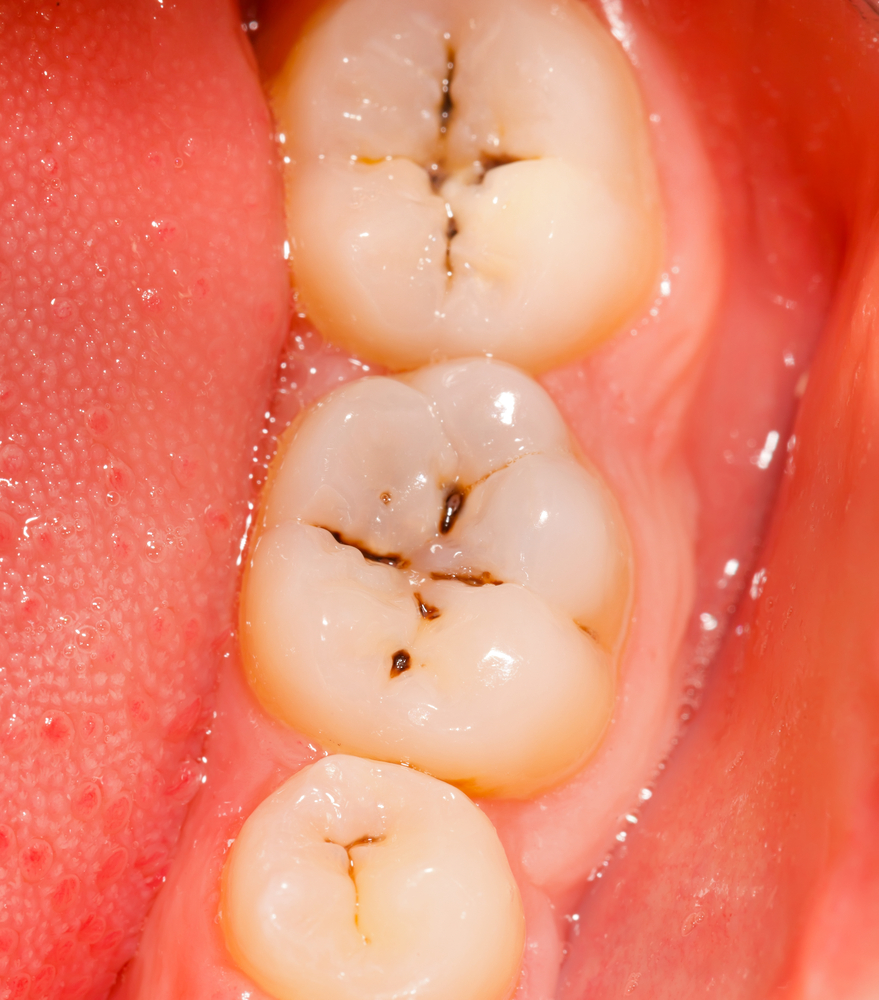

Dental caries is one of the commonest diseases in the world and while a range of adjunctive caries detection devices have been developed visual inspection remains the primary method of detection. However, visual detection can be subjective and examiners can be inconsistent in their interpretation of the clinical appearance of carious lesions. The aim of this review was to determine the overall diagnostic accuracy of visual detection for dental caries in primary and permanent teeth.
Methods
Searches were conducted in the PubMed, Embase, Scopus and OpenSIGLE databases, supplemented by searches of of IADR/ AADR (International and American Associations for Dental Research), and ORCA (European Organisation for Caries Research) congresses for unpublished literature.
Studies involving of visual inspection for detection of primary coronal caries lesions in primary or permanent human teeth with a clearly defined reference standard (Histologic evaluation, operative intervention, direct visual inspection after temporary tooth separation, and radiography) that provided sufficient information to calculate of true positives, false positives, true negatives, false negatives and published in English were considered.
Two reviewers carried out study selections, data abstraction and risk of bias assessment independently. Risk of bias was assessed using the QUADAS-2 checklist. Statistical pooling of sensitivity, specificity, diagnostic odds ratio (DOR), and positive and negative likelihood ratios was carried out using a random effects meta-analysis model, considering different types of settings, teeth, and dental surfaces examined.
Results
- 102 papers published between 1975 and 2014 were included.
- Publication year of included studies ranged from.
- 36% of studies were conducted by experienced examiners;
- 77% were laboratory based;
- 78% were on occlusal surfaces;
- 68% were in permanent teeth;
- 80% used a histologic reference standard
- Pooled sensitivity, specificity and positive likelihood ratios and diagnostic odds ratios (DOR) for the clinical studies only are shown in the tables below:-
| Permanent teeth | No. of studies | Pooled sensitivity (95%CI) | Pooled specificity(95%CI) | Pooled positive likelihood ratio (95%CI) | Pooled diagnostic odds ratio (95%CI) |
| Initial occlusal Caries Lesion |
7 |
0.777(0.755-0.796) | 0.926(0.911-0.939) | 4.649(2.116-10.217) |
30.796 (11.837-80.119) |
| More advanced occlusal caries lesion |
13 |
0.355(0.330-0.382) | 0.918(0.907-0.929) | 4.062(2.404-6.864) |
11.517 (6.783-19.555) |
| Initial proximal Caries Lesion |
3 |
0.297(0.248-0.349) | 0.990(0.985-0.994) | 4.586(0.365-57.589) |
14.039 (0.191-1029.4) |
| More advanced proximal caries lesion |
4 |
0.274(0.191-0.369) | 0.988(0.984-0.992) | 17.947(10.294-31.290) |
23.867 (12.510-45.534) |
| Primary teeth | No. of studies | Pooled sensitivity (95%CI) | Pooled specificity(95%CI) | Pooled positive likelihood ratio (95%CI) | Pooled diagnostic odds ratio (95%CI) |
| Initial occlusal Caries Lesion – |
7 |
0.752(0.708-0.793) | 0.575(0.519-0.626) | 2.678(1.689-4.246) |
11.533 (4.791-27.765) |
| Advanced occlusal caries lesion |
8 |
0.633(0.591-0.730) | 0.881(0.859-0.901) | 4.573(2.444-8.554) |
21.123 (7.909-56.412) |
| Initial proximal Caries Lesion – |
3 |
0.430(0.397-0.464) | 0.908(0.882-0.930) | 2.134(0.972-4.687) |
2.873 (1.087-7.597) |
| More advanced proximal caries lesion |
3 |
0.543 (0.457-0.627) |
0.992 (0.986-0.996) |
46.896(8.882-247.606) |
70.763 (26.979-185.60) |
Conclusions
The authors concluded: –
Visual inspection presents good accuracy in the detection of carious lesions in primary and permanent teeth, with a trend for higher specificity than sensitivity. Importantly, the use of detailed and validated methods improves the accuracy of visual inspection for caries.
Comments
The majority of the studies included in this review are laboratory-based studies an issue raised by the author, and this may have implications for how these findings are translated into clinical practice. They point out that the overall accuracy was similar in the clinical and laboratory based studies although they did note a trend for higher specificities and lower sensitivities in the clinical studies. While higher sensitivities are normally obtained at the expense of reduced specificity higher specificities (ie a lower number of false positives) is considered preferable in a normally slowly progressing disease like caries. The authors recommend that visual examination for caries is used without adjuncts and strongly encourage using a well-established visual scoring system, as this does appear to improve accuracy.
Links
Gimenez T, Piovesan C, Braga MM, Raggio DP, Deery C, Ricketts DN, Ekstrand KR, Mendes FM. Visual Inspection for Caries Detection: A Systematic Review and Meta-analysis. J Dent Res. 2015 May 20. pii: 0022034515586763. [Epub ahead of print] Review. PubMed PMID: 25994176.




Katie Goodman
11 years agoRadiographic caries detection is highly accurate?
11 years agodrjefflo
11 years agoReminovaLtd
11 years agoflyerLPA
11 years agoTheDentalElf
11 years agoTheDentalElf
11 years agoMedwave_cl
11 years agoTheDentalElf
11 years agodrabiddesai
11 years agoHDAssoc
11 years agoTheDentalElf
11 years agofausto_mendes
11 years agoDominic_Hurst
11 years agozmarshman
11 years agoLynette Anear
11 years agoTheDentalElf
11 years agoHumberto Velásquez Mansilla
11 years agoChristine Park
11 years agoHiroshi Kawase
11 years agoToothfairy4you
11 years ago