

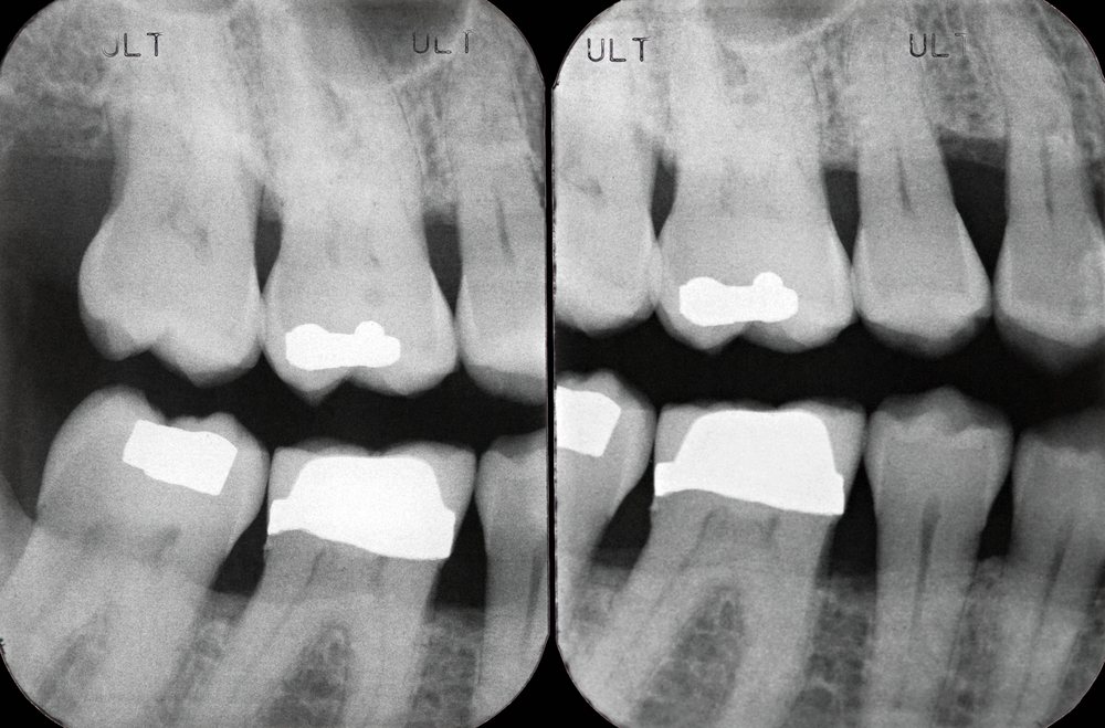

Yesterday we looked at a review of the visual detection of caries. Of the additional aids to caries diagnosis intra-oral radiography is the most frequently used.
The aim of this review was to investigate the accuracy of radiographic caries detection.
Methods
Searches were conducted in the Medline/PubMed, Embase, Cochrane Central, Medion and Open grey databases. Clinical or in vitro studies reporting on intraoral radiography, i.e. bitewing or peri-apical radiographs using a reference standard (gold standard) categorized as (a) destructive or (b) non-destructive (visual-tactile assessment without [occlusal] or with [proximal] tooth separation); destructive methods were further divided into histologic, microradiographic or operative assessment were considered. Only studies providing data allowing construction of a diagnostic 2 x 2 table, i.e. to determine true positive, false negative, false positive and true negative detections were included.
Two independent reviewers assessed studies for inclusion with data from eligible studies being extracted by one reviewer with a 25% independently repeated by a second reviewer. Risk of bias was assessed using the QUADAS-2 tool. Summary measures, sensitivity, specificity and diagnostic odds ratio (DOR) were calculated from the primary dataset. Data synthesis was performed separately for different surfaces (occlusal/proximal) and cut-offs (any kind of lesions, lesions extending into dentine, cavitated lesions) using random-effects meta-analysis.
Results
- 117 studies involving a total 13,375 teeth and 19,108 examined surfaces were included.
- 24 studies were performed in vivo and 93 in vitro.
- The included studies published between 1975 and 2013. All studies were published in English.
- 54 reported on caries detection on occlusal surfaces 55 on proximal surfaces, with 8 reporting on both.
- 92 studies investigated permanent teeth, 23 primary teeth, 2 studies reported on both dentitions.
- Almost all ( 106 studies) performed some kind of destructive validation (histologic or microradiographic analysis or excavation
- 67 studies used analogue radiographs and 25 digital systems.
- 23 were considered to have low risk of bias and 94 a high risk of bias.
- Diagnostic Odds Ratios (DOR) all significantly exceeded 1 (indicating a useful test) and were higher in proximal than occlusal lesions, whilst heterogeneity was moderate (I2 > 50–67%).
| Clinical studies (95% CI) | In vitro studies (95% CI) | |
| Any occlusal caries lesions | ||
| Pooled sensitivity | 0.35 (: 0.31/0.40) | 0.41 (0.39/0.44) |
| Pooled specificity | 0.78 (0.73 – 0.83) | 0.70 (0.76 – 0.84). |
| Diagnostic odds ratio (DOR) | 2.6 (0.80 – 8.2) | 2.4 (1.4 – 3.9) |
| Any kind of proximal lesions | ||
| Pooled sensitivity | 0.24 (0.21 – 0.26) | 0.42 (0.31 – 0.34) |
| Pooled specificity | 0.97 (0.95 – 0.98) | 0.89 (0.88 – 0.90) |
| Diagnostic odds ratio (DOR) | 11.3 (3.9 – 32) | 6.1 (4.6 – 8.22) |
Conclusions
The authors concluded: –
Radiographic caries detection is highly accurate for cavitated proximal lesions, and seems also suitable to detect dentine caries lesions. For detecting initial lesions, more sensitive methods could be considered in population with high caries risk and prevalence.
Comments
As with yesterday’s review of visual inspection the majority of studies included in this review were in vitro and the issues of whether these can be transferred to the clinical situation needs to be considered. The authors consider this in their discussion and they highlight that when comparing the in vitro and clinical findings suggest that the in vitro studies, ‘might over-estimate sensitivity and under-estimate specificity.’
As noted yesterday in caries diagnosis higher specificities (lower number of false positives) is considered preferable in a normally slowly progressing disease like caries. As it is considered that many dentists choose to provide restorative interventions in lesions are confined radiographically to enamel, this the prioritisation of specificity should be emphasised.
Links
Schwendicke F, Tzschoppe M, Paris S. Radiographic caries detection: A systematic review and meta-analysis. J Dent. 2015 Feb 24. pii:S0300-5712(15)00049-4. doi: 10.1016/j.jdent.2015.02.009. [Epub ahead of print]Review. PubMed PMID: 25724114
Dental Elf 9th Jun 2015 – Caries detection: visual detection has good accuracy



Digital imaging for caries: direct digital sensors or photostimulable phosphor plates? - National Elf Service
9 years agojeff_manan
11 years agoHealthwatchTame
11 years agoHWOldham
11 years agoElliot Abt
11 years agoClaudia Cunningham
11 years agoTheDentalElf
11 years agoTheDentalElf
11 years agoTheDentalElf
11 years agoTheDentalElf
11 years agoTheDentalElf
11 years agosergiouribe
11 years agoHumberto Velásquez Mansilla
11 years agoKatie Goodman
11 years agowetherbyortho
11 years agoTheDentalElf
11 years agoChristine Park
11 years agoAdel Azhary
11 years agoCanfordmdvs
11 years agoHiroshi Kawase
11 years ago