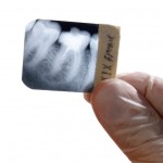
Initially distinguishing between periapical granuloma and a periapical cyst is usually achieved by clinical examination and two-dimensional radiographs, although gold standard standard diagnosis requires histology. However, more advance imaging techniques such as cone- beam computed tomography (CBCT) ultrasonography (US) and magnetic resonance imaging (MRI) have been reported suggesting comparability with histology.
The aim of this review was to assess the diagnostic accuracy of ultrasound in differentiating periapical lesions of endodontic origin in comparison with histopathology.
Methods
Searches were conducted in the PubMed, Scopus, Embase, Web of Science and Pro-Quest databases supplemented by hand searching of the journals, International Endodontic Journal, Journal of Endodontics and Australian Endodontic Journal. English language studies using ultrasound imaging for the differential diagnosis of periapical lesions of endodontic origin in comparison with histopathology were considered. Four reviewers screened and extracted the data with quality assessed using the modified Quality Assessment of Diagnostic Accuracy Studies-2 (QUADAS-2) tool. A random effects model was used for quantitative analysis of the data, and the Deeks test was used for calculating publication bias.
Results
- 12 studies (11 cross-sectional, 1 case report) involving a total sample of 235 patients were included.
- 191 of the 235 patients underwent histological analysis and were included in the review.
- 97 periapical cysts, 90 periapical granulomas and 5 mixed lesions were diagnosed by ultrasound and confirmed by histopathological examination.
- All of the included studies were at high risk of bias and applicability because of concerns over patient selection
- 10 studies contributed to the meta-analysis with summary estimates or sensitivity and specificity of ultrasound to diagnose periapical granulomas and cysts shown in the table.
| Sensitivity (95%CI) | Specificity (95%CI) | |
| Periapical granuloma | 0.94 (0.68 to 0.99) | 0.98 (0.90 to 1.00) |
| Periapical cysts | 0.98 (0.85 to 1.00) | 0.99 (0.71 to 1.00) |
- Publication bias was a concern.
Conclusions
The authors concluded: –
……that the sensitivity and specificity of ultrasound in diagnosing periapical cysts and periapical granulomas were high and accurate. However, taking the quality of evidence into consideration, ultrasound at best can serve as an additional tool for differentiation of cysts and granulomas.
Comments
While 12 studies were included in this review with 10 contributing to the meta-analyses there were concerns regarding the quality of the available evidence. The authors highlighted important issue in relation to patient selection with a lack of clarity regarding patient enrolment in the studies as well as the numbers of observers and blinding. It seems likely that the included samples did not provide a broad spectrum of disease with most patients having established lesions. Most of the lesions (83%) were in the anterior region and there are technical limitations regarding the availability of specially designed ultrasound probes for use in all areas of the mouth. While the review suggests that ultrasound shows promise in this area further well conducted and reported studies of appropriate size and spectrum of disease are required.
Links
Primary Paper
Natanasabapathy V, Arul B, Mishra A, Varghese A, Padmanaban S, Elango S, Arockiam S. Ultrasound imaging for the differential diagnosis of periapical lesions of endodontic origin in comparison with histopathology – a systematic review and meta-analysis. Int Endod J. 2020 Dec 25. doi: 10.1111/iej.13465. Epub ahead of print. PMID: 33368404.
