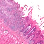
Oral Cancer is currently the 6th most common cancer worldwide. Survival rates have remained largely unchanged for years and this lack of improvement is often attributed to its late presentation. Reducing known risks, smoking, use of tobacco products, alcohol consumption and HPV infection along with early diagnosis are important to improving survival rates.
A range of adjunctive tests have been tried to improve early diagnosis. The aim of this review is to the influence and diagnostic accuracy of these two adjuvant techniques in brush cytology.
Methods
Searches were conducted in the Medline/PubMed, Embase, Elsevier and Web of Science databases Prospective and retrospective studies involving patients with primary, untreated oral precancer where OralCDx brush biopsy or DNA-image cytometry were used to detect disease and histopathology was used as a reference (gold) standard). Only computer assisted methodologies were included.
One reviewer assessed study quality using the quality assessment of diagnostic accuracy studies (QUADAS) checklist. Pooled sensitivity and specificity were calculated with 95% CIs (confidence intervals) separately for each study. Likelihood and diagnostic odds ratios were also calculated along with a summary receiver-operating characteristic (SROC) curve analysis.
Results
- 13 studies involving a total of 1981 oral mucosa lesions were included.
- 8 studies involved OralCDx brush biopsy and 5 DNA-image cytometry.
- Study size was found to be closely related to heterogeneity among studies.
- Analysis suggests a possible publication bias in relation to OralCDx brush biopsy.
| Pooled sensitivity (95%CI) | Pooled specificity (95%CI) | Diagnostic odds ratio (95% CI) | |
| OralCDx brush biopsy | 86%
(81–90%) |
81%
(78–85%) |
20.36
(2.72–152.67) |
| DNA-image cytometry | 89%
(83–94%) |
99%
(97–100%) |
446.08
(73.36–2712.43) |
Conclusions
The authors concluded:
The results of this meta-analysis suggest that DNA-image cytometry has a highly significant potential over OralCDx brush biopsy as an accurate and simple diagnostic tool for clinically suspected oral precancer and oral cancer.
Comments
This review has searched several large databases although only a single author was involved in study selection and data abstraction. Study quality was assessed using the QUADAS tool, which is appropriate for systematic reviews of diagnostic tests and covers 4 key areas, patient selection, the index test, reference standard and the flow and timing of the study.
The authors provide good summary data including pooled sensitivity/specificity, Diagnostic Odds and likelihood ratios. Positive likelihood ratios (+LR) of more than 10 and a negative likelihood ratio (-LR) of below 0.1 are considered to provide strong evidence to rule in or rule out diagnoses respectively in most cases. As diagnostic odds ratio is the ratio of +LR/-LR a figure of more than 100 suggests a good test. Here that average value for DNA-image cytometry is over 400, although the 95% confidence interval is quite wide (73.36–2712.43).
It is also worth noting that the recent Cochrane review by Macey et al looked at diagnostic tests for oral cancer. While the included studies on Oral CDX they did not include studies using DNA-image cytometry. For cytology their meta-analysis included 12 studies and found sensitivity was 0.91 (95%CI; 0.81 to 0.96) and specificity was 0.91 (95%CI; 0.81 to 0.95) with 12 studies included in the meta-analysis. They concluded;
The overall quality of the included studies was poor. None of the adjunctive tests can be recommended as a replacement for the currently used standard of a scalpel biopsy and histological assessment. Given the relatively high values of the summary estimates of sensitivity and specificity for cytology, this would appear to offer the most potential. Combined adjunctive tests involving cytology warrant further investigation.
The discussion of Cochrane review did highlight the potential of DNA-image cytometry and this review provides further evidence that this approach may be of value.
Links
Primary paper
Ye X, Zhang J, Tan Y, Chen G, Zhou G. Meta-analysis of two computer-assisted screening methods for diagnosing oral precancer and cancer. Oral Oncol. 2015 Nov;51(11):966-75. doi:10.1016/j.oraloncology.2015.09.002. Epub 2015 Oct 5. Review. PubMed PMID: 26384539.
Other references
Macey R, Walsh T, Brocklehurst P, Kerr AR, Liu JLY, Lingen MW, Ogden GR, Warnakulasuriya S, Scully C. Diagnostic tests for oral cancer and potentially malignant disorders in patients presenting with clinically evident lesions. Cochrane Database of Systematic Reviews 2015, Issue 5. Art. No.: CD010276. DOI: 10.1002/14651858.CD010276.pub2.
Dental Elf -1st June 2015 – Oral Cancer diagnosis: biopsy and histology still best method
Walsh T, Liu JLY, Brocklehurst P, Glenny AM, Lingen M, Kerr AR, Ogden G, Warnakulasuriya S, Scully C. Clinical assessment to screen for the detection of oral cavity cancer and potentially malignant disorders in apparently healthy adults. Cochrane Database of Systematic Reviews 2013, Issue 11. Art. No.: CD010173. DOI: 10.1002/14651858.CD010173.pub2
Brocklehurst P, Kujan O, O’Malley LA, Ogden G, Shepherd S, Glenny AM. Screening programmes for the early detection and prevention of oral cancer. Cochrane Database of Systematic Reviews 2013, Issue 11. Art. No.: CD004150. DOI: 10.1002/14651858.CD004150.pub4.

DNA-image cytometry and oral cancer diagnosis https://t.co/2kgaOv7Xgy
DNA-image cytometry has potential for oral cancer diagnosis https://t.co/2kgaOv7Xgy
Oral cancer diagnosis-DNA-image cytometry shows potential https://t.co/2kgaOv7Xgy
Oral Cancer – DNA-image cytometry may have promise for early detection https://t.co/2kgaOv7Xgy
DNA-image cytometry may have promise for early oral cancer detection https://t.co/2kgaOv7Xgy
Don’t miss- DNA-image cytometry has potential for oral cancer diagnosis https://t.co/2kgaOv7Xgy